. 2020 Oct 29;17(1):165.
doi: 10.1186/s12985-020-01437-4.

Prediction and evolution of B cell epitopes of surface protein in SARS-CoV-2

Affiliations

Prediction and evolution of B cell epitopes of surface protein in SARS-CoV-2

Jerome Rumdon Lon et al. Virol J. .

Abstract

Background: In order to obtain antibodies that recognize natural proteins, it is possible to predict the antigenic determinants of natural proteins, which are eventually embodied as polypeptides. The polypeptides can be coupled with corresponding vectors to stimulate the immune system to produce corresponding antibodies, which is also a simple and effective vaccine development method. The discovery of epitopes is helpful to the development of SARS-CoV-2 vaccine.

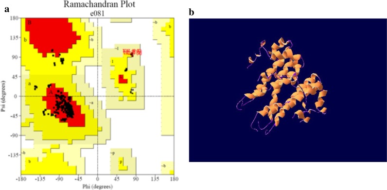
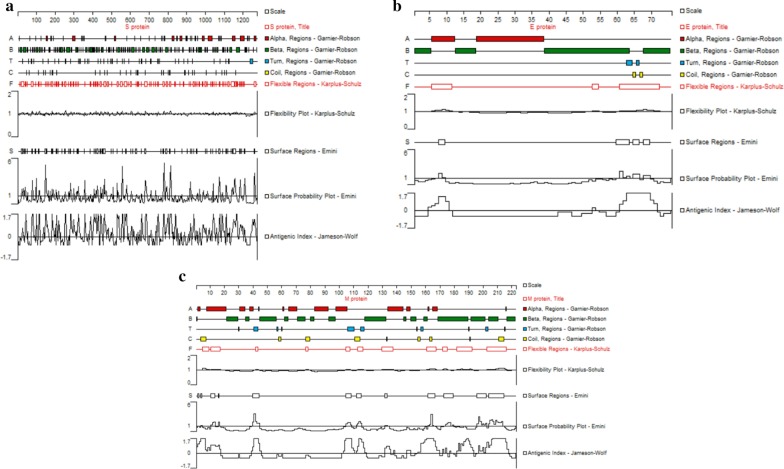
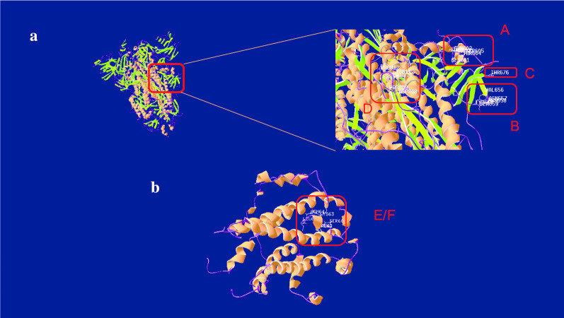
Methods: The analyses were related to epitopes on 3 proteins, including spike (S), envelope (E) and membrane (M) proteins, which are located on the lipid envelope of the SARS-CoV-2. Based on the NCBI Reference Sequence: NC_045512.2, the conformational and linear B cell epitopes of the surface protein were predicted separately by various prediction methods. Furthermore, the conservation of the epitopes, the adaptability and other evolutionary characteristics were also analyzed, the sequences of the whole genome of SARS-CoV-2 were obtained from the GISAID.

Results: 7 epitopes were predicted, including 6 linear epitopes and 1 conformational epitope. One of the linear and one of the conformational consist of identical sequence, but represent different forms of epitopes. It is worth mentioning that all 6 identified epitopes were conserved in nearly 3500 SARS-CoV-2 genomes, showing that it is helpful to obtain stable and long-acting epitopes under the condition of high frequency of amino acid mutation, which deserved further study at the experiment level.

Conclusion: The findings would facilitate the vaccine development, had the potential to be directly applied on the prevention in this disease, but also have the potential to prevent the possible threats caused by other types of coronavirus.

Keywords: Bioinformatics; Epitopes; Evolution; SARS-CoV-2.


Conflict of interest statement

The authors declare that they have no competing interests.

Figures

Fig. 1
Fig. 1
The 3D structure prediction and Ramachandran plot analysis of the E protein. a The Ramachandran plot analysis of the 3D structure of the E protein (without Gly and Pro). All of the residues located on the allowed region. indicating that the structure was reliable from a thermodynamic point of view. b The 3D structure of the E protein predicted by homology modeling. It is a pentamer with ion channel activity [38]. Its head is short, the middle of the tail is a transmembrane region which help the E protein embed in the envelope of SARS-CoV-2
Fig. 2
Fig. 2
The secondary structures and properties analysis of the S, E and M protein. a Analysis of the S protein. It contains most α-helix and β-sheet, some Turn and Coli region, several discontinuous high flexibility fragments, fluctuant surface probability with a few of positive peak and several antigenicity regions with positive peak. The S protein showed concentrated high antigenicity peaks in 600–800 residues. b Analysis of the E protein. It contains most α-helix and β-sheet, some Turn and Coli region, three high flexibility fragments, few surface probability regions and two antigenicity regions with positive peak in the begin and the end of polypeptide chain, respectively. The E protein showed concentrated high antigenicity peaks in 60–70 residues. c Analysis of the M protein. It contains most α-helix and β-sheet, some Turn and Coli region, several high flexibility fragments, few surface probability regions, two antigenicity region with positive single peak in the begin and middle of peptide chain, respectively, and consecutive positive peaks in the end. The M protein showed concentrated high antigenicity peaks in 200–220 residues. Interestingly, the high antigenicity peaks of all three proteins were in the region where the α-helix is relatively sparse, which may be related to the fact that the α-helix structure of the helix prevents continuous residues from being located on the surface
Fig. 3
Fig. 3
The predicted epitopes of the S and E protein. a The predicted linear B-cell epitopes of the S protein. The epitope A, B, C located in the forepart of the tail, the epitope D located in the back part of the tail and is close to the transmembrane region. b The predicted B-cell epitope of the E protein. The epitope G is the linear epitope and the F is the conformational epitope, which are coincide

References

    1. Han Q, Lin Q, Jin S, You L. Coronavirus 2019-nCoV: a brief perspective from the front line. J Infect. 2020;80:373–377. doi: 10.1016/j.jinf.2020.02.010. - DOI - PMC - PubMed
    1. Ying L, Gayle AA, Annelies WS, Joacim R. The reproductive number of COVID-19 is higher compared to SARS coronavirus. J Travel Med. 2020. - PMC - PubMed
    1. El-Manzalawy Y, Honavar V. Recent advances in B-cell epitope prediction methods. Immunome Res. 2010;6(Suppl 2):S2. doi: 10.1186/1745-7580-6-S2-S2. - DOI - PMC - PubMed
    1. Sun P, Ju H, Liu Z, Ning Q, Zhang J, Zhao X, Huang Y, Ma Z, Li Y. Bioinformatics resources and tools for conformational B-cell epitope prediction. Comput Math Methods Med. 2013;2013:943636. - PMC - PubMed
    1. Bai Y, Jiang D, Lon JR, Chen X, Hu M, Lin S, Chen Z, Wang X, Meng Y, Du H. Comprehensive evolution and molecular characteristics of a large number of SARS-CoV-2 genomes reveal its epidemic trends. Int J Infect Dis. 2020;100:164–173. doi: 10.1016/j.ijid.2020.08.066. - DOI - PMC - PubMed

Publication types

MeSH terms

LinkOut - more resources