Review
. 2021 Aug:163:14-22.
doi: 10.1016/j.pbiomolbio.2020.10.007. Epub 2020 Oct 24.

The dynamic nature of the Mre11-Rad50 DNA break repair complex

Affiliations
Review

The dynamic nature of the Mre11-Rad50 DNA break repair complex

Mahtab Beikzadeh et al. Prog Biophys Mol Biol. 2021 Aug.

Abstract

The Mre11-Rad50-Nbs1/Xrs2 protein complex plays a pivotal role in the detection and repair of DNA double strand breaks. Through traditional and emerging structural biology techniques, various functional structural states of this complex have been visualized; however, relatively little is known about the transitions between these states. Indeed, it is these structural transitions that are important for Mre11-Rad50-mediated DNA unwinding at a break and the activation of downstream repair signaling events. Here, we present a brief overview of the current understanding of the structure of the core Mre11-Rad50 complex. We then highlight our recent studies emphasizing the contributions of solution state NMR spectroscopy and other biophysical techniques in providing insight into the structures and dynamics associated with Mre11-Rad50 functions.

Keywords: Allostery; DNA double Strand break repair; DNA repair; Methyl TROSY; Mre11-Rad50-Nbs1; NMR spectroscopy.


Figures

Fig. 1.
Fig. 1.
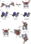
Domain motions that are critical for Mre11-Rad50 function. A) X-ray crystal structures of T. maritima MR complex in the ATP-free ‘open’ (left; PDB entry 3QG5) and ATP-bound ‘closed’ (right; PDB entry 3THO) conformations (Lammens et al., 2011; Möckel et al., 2012). For each structure, Mre11 nuclease, capping, and helix-loop-helix domains are colored light blue, grey, and purple, respectively, while Rad50 NBD N- and C-terminal sub-domains are colored red and gold. B) X-ray crystal structures of P. furiosus Mre11 capping and nuclease domain (PDB entries 3DSC and 3DSD) with nuclease and capping domains colored in blue and grey shades, respectively (Williams et al., 2008). Catalytic Mn2+ ions are denoted by magenta circles. The effect of asymmetric domain motions is shown on the cartoon DNA. C) Cryo-EM structures of E. coli MR (SbcCD) complex in the ATP-bound ‘resting’ state (left; PDB entry 6S6V) and ADP- and dsDNA-bound ‘cutting’ state (right; PDB entry 6S85). Structures are colored as in A. Three views of the ‘cutting’ state are presented where the structure has been rotated by the indicated angle.
Fig. 2.
Fig. 2.
Small- and large-scale conformational motions within Rad50 NBDs. A) A region of the methyl-TROSY spectra for wild type and basic switch/hinge (left) and D-loop (right) mutants highlighting changes in structure that occur upon perturbation. B) Schematic of the structural changes occurring in the Rad50 NBDs during ATP-induced association, hydrolysis, and dissociation. Red and gold shapes symbolize the N- and C-terminal sub-domains of the NBDs, respectively, and the changes in the shapes signify the different states the NBDs must populate to associate, hydrolyze ATP, and dissociate. The pentagons and stars represent ATP, whereas the circles represent ADP. Basic switch and hinge region mutants accelerate the Rad50NBD association step, whereas D-loop mutants accelerate cooperative/allosteric transitions leading to faster ATP hydrolysis. C) An example of LRET-derived distance distribution demonstrating a ‘partially open’ state. ATP binding shifts the equilibrium to favor the ‘closed’ state and leads to slightly shorter distance between the two probes for both states (dashed line). D) Cartoon representation of the new ‘partially open’ state of the MR complex observed in LRET experiments. Domains of the MR complex are colored according to Figure 1A. Figure adapted from Refs. (Boswell et al., 2020, 2018).
Fig. 3.
Fig. 3.
Rigid Mre11 recognizes dsDNA substrates for exonuclease activity. A) Overlay of the methyl-TROSY spectra indicating that dsDNA and ssDNA substrates cause a single and unique perturbation. B) Overlay of the methyl-TROSY spectra showing that the separation-of-nuclease function mutants (Pf Mre11ND Y187C and H52S) bind dsDNA (left) differently compared to wild type but ssDNA (right) the same. C) Example methyl 1H-1H dipolar cross-correlated triple quantum relaxation data (Sun et al., 2011) for wild type, Y187C, and H52S Pf Mre11ND. The extracted relaxation rate (η) reports on the amplitude of side-chain methyl group dynamics as schematically shown to the right. D) A cartoon representation of a less flexible Mre11ND that can stably bind and recognize dsDNA and ssDNA substrates for nuclease reactions. Separation-of-nuclease function mutants alter the structure and dynamics of the protein leading to the inability to properly recognize dsDNA substrates. Figure adapted from Ref. (Rahman et al., 2020a).

References

    1. Al-Ahmadie H, Iyer G, Hohl M, Asthana S, Inagaki A, Schultz N, Hanrahan AJ, Scott SN, Brannon AR, McDermott GC, Pirun M, Ostrovnaya I, Kim P, Socci ND, Viale A, Schwartz GK, Reuter V, Bochner BH, Rosenberg JE, Bajorin DF, Berger MF, Petrini JHJ, Solit DB, Taylor BS, 2014. Synthetic Lethality in ATM-Deficient RAD50-Mutant Tumors Underlies Outlier Response to Cancer Therapy. Cancer Discov. 4, 1014–1021. 10.1158/2159-8290.CD-14-0380 - DOI - PMC - PubMed
    1. Boehr DD, Nussinov R, Wright PE, 2009. The role of dynamic conformational ensembles in biomolecular recognition. Nat. Chem. Biol 5, 789–796. 10.1038/nchembio.232 - DOI - PMC - PubMed
    1. Boswell ZK, Canny MD, Buschmann TA, Sang J, Latham MP, 2020. Adjacent mutations in the archaeal Rad50 ABC ATPase D-loop disrupt allosteric regulation of ATP hydrolysis through different mechanisms. Nucleic Acids Res 48, 2457–2472. 10.1093/nar/gkz1228 - DOI - PMC - PubMed
    1. Boswell ZK, Latham MP, 2019. Methyl-Based NMR Spectroscopy Methods for Uncovering Structural Dynamics in Large Proteins and Protein Complexes. Biochemistry 58, 144–155. 10.1021/acs.biochem.8b00953 - DOI - PMC - PubMed
    1. Boswell ZK, Rahman S, Canny MD, Latham MP, 2018. A dynamic allosteric pathway underlies Rad50 ABC ATPase function in DNA repair. Sci. Rep 8, 1–12. 10.1038/s41598-018-19908-8 - DOI - PMC - PubMed

Publication types

MeSH terms