Review
. 2020 Dec:201:108330.
doi: 10.1016/j.exer.2020.108330. Epub 2020 Oct 27.

USH2A-retinopathy: From genetics to therapeutics

Affiliations
Review

USH2A-retinopathy: From genetics to therapeutics

Lyes Toualbi et al. Exp Eye Res. 2020 Dec.

Abstract

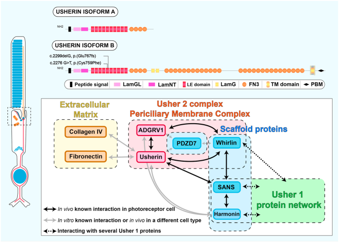
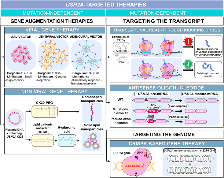
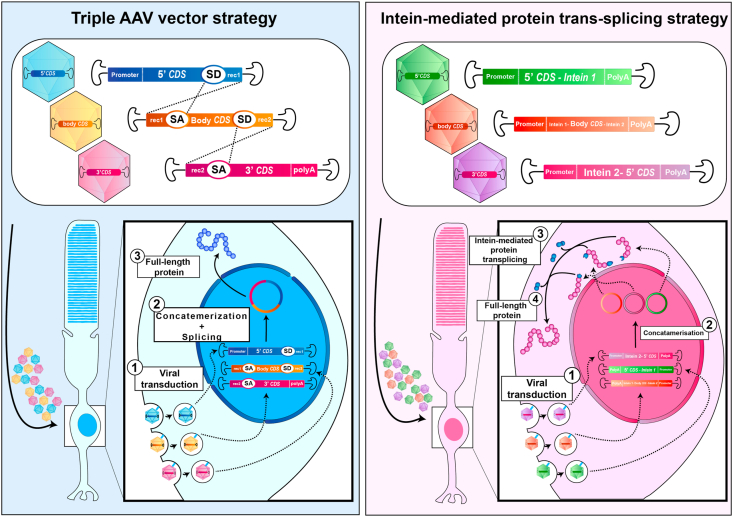
Bilallelic variants in the USH2A gene can cause Usher syndrome type 2 and non-syndromic retinitis pigmentosa. In both disorders, the retinal phenotype involves progressive rod photoreceptor loss resulting in nyctalopia and a constricted visual field, followed by subsequent cone degeneration, leading to the loss of central vision and severe visual impairment. The USH2A gene raises many challenges for researchers and clinicians due to a broad spectrum of mutations, a large gene size hampering gene therapy development and limited knowledge on its pathogenicity. Patients with Usher type 2 may benefit from hearing aids or cochlear implants to correct their hearing defects, but there are currently no approved treatments available for the USH2A-retinopathy. Several treatment strategies, including antisense oligonucleotides and translational readthrough inducing drugs, have shown therapeutic promise in preclinical studies. Further understanding of the pathogenesis and natural history of USH2A-related disorders is required to develop innovative treatments and design clinical trials based on reliable outcome measures. The present review will discuss the current knowledge about USH2A, the emerging therapeutics and existing challenges.

Keywords: Disease models; Hair cells; Photoreceptor; Retinitis pigmentosa; Therapy; USH2A; Usher syndrome; Usherin.


Conflict of interest statement

The authors declare no competing interests.

Figures

Fig. 1
Fig. 1
Diagram summarizing the prevalence of the Usher syndrome subtypes and USH2A pathogenic variants.
Fig. 2
Fig. 2
Schematic diagram of usherin localisation in photoreceptors and hair cells. (A) Cellular organisation of a photoreceptor. The photoreceptor possesses an inner segment and an outer segment, a highly specialised cilium responsible for light detection. The inner segment is connected to the outer segment through the connecting cilium. (B) The connecting cilium is wrapped in the periciliary membrane complex (blue), where the usherin long isoform (red) is spatially restricted. (C) Side view and section (D) of the periciliary membrane complex. (E) Hair cells are the ciliated sensory cells of the cochlea responsible for the transformation of sound-induced vibrations into electric signals. Usherin (red) is localised to the ankle link of developing post-natal hair cells. (For interpretation of the references to colour in this figure legend, the reader is referred to the web version of this article.)
Fig. 3
Fig. 3
Usherin isoforms and interacting partners in photoreceptors. Usherin isoform a consists of 1 LamG-like jellyroll fold domain (LamGL), 1 Laminin N-terminal domain (LamNT), 10 laminin EGF-like domains (LE domain) and 4 fibronectin type III repeats. In addition to these domains, usherin isoform b is composed of 2 laminin G domains (LamG), 28 fibronectin type III repeats (FN3), a transmembrane domain (TM domain) and an intracellular PDZ-binding domain (PBM). The 2 most common mutations (c.2299delG, p.Glu767Serfs*21 and c2276 G>T, p.Cys759Phe) are located in the 5th laminin domain. Interacting partners have been divided into 4 groups: the Usher 2 complex periciliary membrane complex (red), the extracellular matrix partners (yellow), the scaffold proteins (blue) and the Usher 1 protein network (green). (For interpretation of the references to colour in this figure legend, the reader is referred to the web version of this article.)
Fig. 4
Fig. 4
Therapeutic strategies for USH2A-related disease.USH2A-targeted strategies can be divided into two categories, mutation-independent and -dependent. The mutation-independent can be applied to all the USH2A-related diseases regardless of the pathogenic mutation. It involves the generation of healthy copies of USH2A coding sequence (CDS) using viral or non-viral vectors. AAV and lentiviral vectors cannot accommodate the USH2A CDS while adenoviral vectors can. Mutation-dependent strategies have to be applied to a specific mutation or type of mutation. It can target the transcript using translational read-through inducing drugs (e.g. nonsense mutation) and antisense oligonucleotides (e.g. pseudo-exon inclusion), or target the genome using CRISPR-Cas9 editing (e.g. missense mutation). AAV, adeno-associated vector; CK30-PEG, 30-mer polylysine conjugated with polyethylene glycol; DOTAP, 1,2-dioleoyl-3-trimethylammonium-propane; TRIDs, translational read-through inducing drugs; NMD, nonsense-mediated decay.
Fig. 5
Fig. 5
Expanding AAV vector capacity for large gene transfer. The scheme represents two strategies that allow the successful transfer of large genes. The first strategy on the left panel is based on the concatemerisation and splicing of three transgenes to reconstitute the full-length coding sequence of interest. AAV vectors carrying the three different transgenes transduce the photoreceptor cell. The transgenes concatemerise and splice into a single episome in the nucleus allowing the production of the full-length protein of interest. The second strategy consists of intein-mediated transplicing allowing the reconstitution of the full-length protein. Similarly to the previous strategy, AAV vectors carrying three different transgenes transduce the targeted cell. However, it forms three distinct episomes. The three proteins resulting from these episomes by intein-mediated transplicing, lead to the full-length protein. AAV, adeno-associated virus; CDS, coding sequence; SD, splicing donor; SA, splicing acceptor; rec, recombinogenic region.

References

    1. Abadie C., Blanchet C., Baux D., Larrieu L., Besnard T., Ravel P., Biboulet R., Hamel C., Malcolm S., Mondain M., Claustres M., Roux A.-F. Audiological findings in 100 USH2 patients. Clin. Genet. 2012;82:433–438. doi: 10.1111/j.1399-0004.2011.01772.x. - DOI - PubMed
    1. Adato A., Lefèvre G., Delprat B., Michel V., Michalski N., Chardenoux S., Weil D., El-Amraoui A., Petit C. Usherin, the defective protein in Usher syndrome type IIA, is likely to be a component of interstereocilia ankle links in the inner ear sensory cells. Hum. Mol. Genet. 2005;14:3921–3932. doi: 10.1093/hmg/ddi416. - DOI - PubMed
    1. Ahmed Z.M., Jaworek T.J., Sarangdhar G.N., Zheng L., Gul K., Khan S.N., Friedman T.B., Sisk R.A., Bartles J.R., Riazuddin Sheikh, Riazuddin Saima. Inframe deletion of human ESPN is associated with deafness, vestibulopathy and vision impairment. J. Med. Genet. 2018;55:479–488. doi: 10.1136/jmedgenet-2017-105221. - DOI - PMC - PubMed
    1. Aller E., Larrieu L., Jaijo T., Baux D., Espinós C., González-Candelas F., Nájera C., Palau F., Claustres M., Roux A.-F., Millán J.M. The USH2A c.2299delG mutation: dating its common origin in a Southern European population. Eur. J. Hum. Genet. 2010;18:788–793. doi: 10.1038/ejhg.2010.14. - DOI - PMC - PubMed
    1. Angueyra J.M., Kindt K.S. Leveraging zebrafish to study retinal degenerations. Front. Cell Dev. Biol. 2018 doi: 10.3389/fcell.2018.00110. - DOI - PMC - PubMed

Publication types