. 2020 Oct 29;3(12):e202000742.
doi: 10.26508/lsa.202000742. Print 2020 Dec.

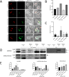
Leishmania-infected macrophages release extracellular vesicles that can promote lesion development

Affiliations

Leishmania-infected macrophages release extracellular vesicles that can promote lesion development

Anna Gioseffi et al. Life Sci Alliance. .

Abstract

Leishmania donovani infection of macrophages results in quantitative and qualitative changes in the protein profile of extracellular vesicles (EVs) released by the infected host cells. We confirmed mass spectrometry results orthogonally by performing Western blots for several Leishmania-infected macrophage-enriched EVs (LieEVs) molecules. Several host cell proteins in LieEVs have been implicated in promoting vascular changes in other systems. We also identified 59 parasite-derived proteins in LieEVs, including a putative L. donovani homolog of mammalian vasohibins (LdVash), which in mammals promotes angiogenesis. We developed a transgenic parasite that expressed an endogenously tagged LdVash/mNeonGreen (mNG) and confirmed that LdVash/mNG is indeed expressed in infected macrophages and in LieEVs. We further observed that LieEVs induce endothelial cells to release angiogenesis promoting mediators including IL-8, G-CSF/CSF-3, and VEGF-A. In addition, LieEVs induce epithelial cell migration and tube formation by endothelial cells in surrogate angiogenesis assays. Taken together, these studies show that Leishmania infection alters the composition of EVs from infected cells and suggest that LieEVs may play a role in the promotion of vascularization of Leishmania infections.


Conflict of interest statement

The authors declare that they have no conflict of interest.

Figures
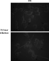
Figure S1.
Figure S1.. Imaging of RAW264.7 macrophages 72 h postinfection.
Macrophage infection cultures were thoroughly washed after 24 h. After an additional 48 h of culture, the culture medium was recovered for isolation of EVs. Imaging analysis was performed to confirm absence of external parasites. Representative bright-field images of RAW264.7 macrophages 72 h postinfection indicated that few external parasites remained after washing the plates at 24 h. Images were acquired on AxioObserver microscope using differential inference contrast microscopy and 15 z-stack images were taken at 0.3-μm intervals for each frame. Image files were processed using Fiji and final images represent 4 z slices combined. Arrows represent infected macrophages. Cultures are evaluated after each EV isolation; however, these images are representative of three recent biological repeats.
Figure 1.
Figure 1.. Isolation and characterization of extracellular vesicles from Leishmania donovani–infected macrophages.
(A) Workflow for collection of extracellular vesicles from RAW264.7 macrophages infected with L. donovani parasites. Infection was performed in media supplemented with exosome-depleted serum. After 24 h, the culture medium was removed. Cultures were washed to remove uninternalized parasites and replenished with fresh medium supplemented with exosome-depleted serum. After an additional 48 h, culture medium was recovered, pooled, and processed following the centrifugation and filtration steps shown in the figure. (B) Nanoparticle tracking analysis was performed from which particle size distribution and particle concentration was obtained. Plot of particles/cell was calculated using cell count at the end of the infection. Data for graphs were obtained from multiple experiments (ceEV n = 7, LieEV n = 7; *P = 0.0082). (C) Representative image of vesicles in LieEV preparation processed for scanning electron microscopy. (D) Representative transmission electron microscopy image of immunogold CD9-labeled particles in LieEVs. Arrows point to gold particles denoting reactivity of antibody.
Figure 2.
Figure 2.. Mouse derived molecules in the LieEV proteome, includes known exosome markers as well as molecules implicated in mediating vascular changes in other systems.
The proteome of LieEVs recovered after 72-h infection was compared with the proteome of ceEVs from uninfected cells. (A) Venn diagram was plotted using proteins identified by mass spectrometry and partitioned according to sample type. The presence and absence of host molecules with and without infection and common to both samples are indicated. (B) The protein content of the preparations was revealed by Ponceau S staining of the blots to normalize for material loaded into each well for each sample type. Approximately 1 × 10¹⁰ particles from EV preparations from three replicate experiments and 50 μg of lysates from infected cells at the 72 h infection point were analyzed. (C) Western blot was performed to confirm the presence of known and novel exosome markers. Uninfected macrophages were treated in an identical manner as infected samples. The blots were then probed with anti-CD9, stripped, and probed with anti-Annexin A3. Identical blots were probed initially with anti-CD63, stripped, and probed with anti-calnexin. (D) Quantification was performed by measuring the mean gray background area using ImageJ software. Background pixel density was subtracted from the inverse of each measurement to obtain relative quantification values. Analysis of the blots showed that CD9 was significantly more abundant in LieEVs than ceEVs and cell lysates (*P = 0.0261, n = 3), whereas levels of CD63 were comparable for all samples. Annexin A3 was significantly more abundant in LieEVs than in ceEVs (*P = 0.0123, n = 3). Calnexin was barely detected in EVs as compared with cell lysates. Statistical test for differences was by ANOVA. Source data are available for this figure.
Figure 3.
Figure 3.. Analysis of the host proteome by Ingenuity Pathway Analysis suggested that murine host proteins in LieEVs may differentially affect biological pathways and functions.
(A) Subset of LieEV molecules classified in the top 5 “Molecular and Cellular Functions,” top 5 “Diseases and Disorders” and top five Physiological systems development and function. (B) Most of the molecules in LieEVs that were categorized under cardiovascular system development have been implicated in the control of angiogenesis. Only those proteins that had a fold change >2 and P-value < 0.05 were analyzed. Canonical pathways were determined by using a right-tailed Fisher exact test by ingenuity pathway analysis.
Figure S2.
Figure S2.. The open reading frame of the Leishmania vasohibin homolog encodes a longer protein as compared with the two mouse vasohibin molecules.
(A) The predicted amino acid sequence of LdVash (A). Shaded region shows peptide to which an antiserum was generated. (B) Alignment of LdVash with mouse Vash 1 and Vash 2.
Figure S3.
Figure S3.. Scheme for endogenous tagging of LdVash.
(A) The puromycin gene and 2A peptide along with the mNeonGreen gene were fused in-frame to the endogenous LdVash gene via homologous recombination (A). Recombinant parasites were selected by growth in puromycin. Upon expression, there is co-translational cleavage via the 19–amino acid 2A peptide sequence that separates the LdVash/mNG protein from the puromycin protein. (B) For verification of appropriate integration, the 5′ prime primer was generated from the 3′ region of the puromycin gene, whereas the 3′ primer was from the 3′ region of the LdVash gene. Insertion at the predicted integration site would be expected to result in the amplification of a 1,255-bp product. (C) Agarose gel image showing the amplification product obtained from the L9 recombinant line.
Figure 4.
Figure 4.. The parasite homolog of vasohibin is expressed in Leishmania-infected cells and is a cargo in EVs from infected cells.
Two parasites lines were derived in which the LdVash gene was endogenously tagged with mNeonGreen (LdVash/mNG+) (FL3 and L9). RAW264.7 macrophages were infected with the LdVash/mNG+ lines and analyzed for the distribution of mNG in infected cells and in EVs. (A) Representative images obtained from live infected cells at the indicated times. Metacyclic FL3 parasites were incubated with CMPTX-red before initiation of infection. White arrows point to parasites in the infected cells. Green label shows the distribution of LdVash/mNG as the infection progresses. These figures are representative of at least four live cell imaging experiments. (B) Particle analysis by NanoSight tracking analysis of total EVs recovered from uninfected cells or from cells infected with either of the two LdVash/mNG+ parasite lines at indicated times. (B, C) Enumeration of fluorescent EV particles by NanoSight tracking analysis in the samples in (B). Data in (B) and (C) were compiled from 1 representative of three experiments. Increases in EVs released by older infections was significant (by ANOVA); however, presence of fluorescent EVs was reproducible but not statistically significant. (D) Western blot analysis of lysates and EVs from LdVash/mNG+ infected cells. Blots were probed with antiserum to LdVash and anti-mNG. Blots are representative of three experiments. (E) Densitometric scans of indicated bands from three experiments are shown. Statistical test for differences was by ANOVA. Source data are available for this figure.
Figure S4.
Figure S4.. Expression of LdVash/mNG in primary macrophages.
The recombinant lines were used to infect primary macrophages in peritoneal exudate cells. Metacyclic parasites from the L9 LdVash/nNG line were incubated with macrophages. (A) After washing off uninternalized parasites, live cell images were captured at the indicated times. The arrows point to parasites within macrophages, which was confirmed with the bright field image. (B) After infection of primary cells for 4 h, parasites were washed off, and the medium was replaced with DMEM supplemented with exosome-depleted serum. EVs were prepared from 24-h-old infections or from 96-h-old infections. NanoSight tracking analysis was performed to enumerate the total number of EVs at either 24 or 96 h. The proportion of LieEV particles with fluorescence was also determined. The data were compiled from two experiments. Reduction in fluorescent LieEVs at 96 h is likely due to death of recombinant parasites in primary exudate cells.
Figure S5.
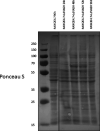
Figure S5.. Ponceau S staining of Western blot for analysis of LdVash and LdVash/mNG expression in infected cells.
Lysates and EVs from LdVash/mNG+ infected cells were analyzed for expression of LdVash and LdVash/mNG. This figure is a representative Ponceau S stain showing equal loading of samples analyzed in Fig 4D. Source data are available for this figure.
Figure 5.
Figure 5.. LieEVs promote endothelial cell tube formation and cell migration.
EVs from infected cells were evaluated alongside EVs from uninfected cells in surrogate assays of angiogenesis. (A) Representative images of gap closure at indicated times by the MDA-MB-231 epithelial cell in a scratch assay are shown. (B) Migration after incubation with intact LieEVs and ceEVs was captured over a period of 22 h and plotted. (C) Migration was also evaluated after incubation with disrupted LieEVs and ceEVs. Data were compiled from two biological repeats. (D) Capacity of LieEVs and ceEVs to promote tube formation by the HUVEC line was determined after incubation for 6 h. A representative image shows that intact LieEVs induce well organized tubes, which contrasts with either disrupted LieEVs or intact or disrupted ceEVs. (E) Changes in tube length and number of branching points induced by treatment with VEGF, LieEVs and ceEVs were plotted. Disrupted LieEVs and ceEVs and suramin that inhibits endothelial cell movement were also analyzed. For statistical analysis of differences, one-way ANOVA was performed with multiple comparisons for each time point from at least two biological repeats, followed by Brown–Forsythe’s and Bartlett’s tests.
Figure 6.
Figure 6.. LieEVs activate endothelial cells to release angiogenesis promoting cytokines.
A multianalyte assay of secreted molecules implicated in angiogenesis was performed on supernatant fluid recovered after 24-h incubation of LieEVs or ceEVs with endothelial cells. Concentrations of each analyte were calculated from standard curves that were included in each experiment. Data were compiled from two biological repeats and analyzed using one-way ANOVA with multiple comparisons for statistical analysis.

References

    1. Abels ER, Breakefield XO (2016) Introduction to extracellular vesicles: Biogenesis, RNA cargo selection, content, release, and uptake. Cell Mol Neurobiol 36: 301–312. 10.1007/s10571-016-0366-z - DOI - PMC - PubMed
    1. Aillaud C, Bosc C, Peris L, Bosson A, Heemeryck P, Van Dijk J, Le Friec J, Boulan B, Vossier F, Sanman L, et al. (2017) Vasohibins/SVBP are tubulin carboxypeptidases (TCPs) that regulate neuron differentiation. Science 358: 1448–1453. 10.1126/science.aao4165 - DOI - PubMed
    1. Akers JC, Gonda D, Kim R, Carter BS, Chen CC (2013) Biogenesis of extracellular vesicles (EV): Exosomes, microvesicles, retrovirus-like vesicles, and apoptotic bodies. J Neurooncol 113: 1–11. 10.1007/s11060-013-1084-8 - DOI - PMC - PubMed
    1. Atayde V, Hassani K, da Silva Lira Filho A, Borges A, Adhikari A, Martel C, Olivier M (2016) Leishmania exosomes and other virulence factors: Impact on innate immune response and macrophage functions. Cell Immunol 309: 7–18. 10.1016/j.cellimm.2016.07.013 - DOI - PubMed
    1. Bachurski D, Schuldner M, Nguyen PH, Malz A, Reiners KS, Grenzi PC, Babatz F, Schauss AC, Hansen HP, Hallek M, et al. (2019) Extracellular vesicle measurements with nanoparticle tracking analysis: An accuracy and repeatability comparison between NanoSight NS300 and ZetaV iew. J Extracell Vesicles 8: 1596016 10.1080/20013078.2019.1596016 - DOI - PMC - PubMed

Publication types