Case Reports
. 2020 Oct 29;13(10):e233148.
doi: 10.1136/bcr-2019-233148.

Traumatic avulsion of the globe with optic nerve transection: an unusual presentation

Affiliations
Case Reports

Traumatic avulsion of the globe with optic nerve transection: an unusual presentation

Seema Meena et al. BMJ Case Rep. .

Abstract

Complete globe extrusion, whether traumatic or spontaneous, is a rare clinical entity and if associated with optic nerve avulsion, it has a worse visual outcome, though repositioning of the globe may be attempted. We report a case of road traffic accident, wherein the patient presented with an extrusion of the globe, along with a complete transection of the optic nerve, about 4 cm from the optic nerve head, with only a residual attachment to the orbital rim via the unsevered lateral conjunctival flap, where the enucleation was completed and the conjunctiva was sutured.

Keywords: ophthalmology; visual pathway.


Conflict of interest statement

Competing interests: None declared.

Figures
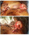
Figure 1
Figure 1
(A) Clinical photograph of the patient at presentation to the casualty unit, showing sutured facial lacerations and an avulsed left globe. (B) Clinical photograph of the patient, showing an avulsed left globe with completely transected optic nerve stump measuring approximately 4 cm.
Figure 2
Figure 2
Non-contrast CT of head and orbit axial view depicting an extrusion of left globe with an attached transected optic nerve stump. Globe is attached to the orbital rim temporally via the lateral rectus muscle. Avulsed medial rectus muscle and multiple fractures of medial wall of orbit and nasal septum can be appreciated.
Figure 3
Figure 3
Clinical photograph of the patient at 2 weeks post-surgery, showing sutured facial and right upper eyelid laceration and left post-enucleated socket.

References

    1. Bajaj MS, Kedar S, Sethi A, et al. . Traumatic globe luxation with optic nerve transection. Orbit 2000;19:165–70. 10.1076/orbi.19.3.165.2661 - DOI - PubMed
    1. Unal S, Argin A, Arslan E, et al. . Bilateral complete avulsion of ocular globes in a Le Fort III maxillofacial fracture: a case report and review of the literature. Eur J Ophthalmol 2005;15:123–5. 10.1177/112067210501500119 - DOI - PubMed
    1. Tok L, Tok OY, Argun TC, et al. . Bilateral traumatic globe luxation with optic nerve transection. Case Rep Ophthalmol 2014;5:429–34. 10.1159/000370043 - DOI - PMC - PubMed
    1. Roki N, Roka YB, Acharya R. Traumatic avulsion and bilateral eye loss: report of two cases. Nepal J Ophthalmol 2013;5:271–4. 10.3126/nepjoph.v5i2.8743 - DOI - PubMed
    1. Lang GK, Bialasiewicz AA, Röhr WD. [Bilateral traumatic eye avulsion]. Klin Monbl Augenheilkd 1991;198:112–6. 10.1055/s-2008-1045940 - DOI - PubMed

Publication types