. 2020 Nov 17;117(46):29090-29100.
doi: 10.1073/pnas.2001177117. Epub 2020 Oct 29.

The structural basis for an on-off switch controlling Gβγ-mediated inhibition of TRPM3 channels

Affiliations

The structural basis for an on-off switch controlling Gβγ-mediated inhibition of TRPM3 channels

Marc Behrendt et al. Proc Natl Acad Sci U S A. .

Abstract

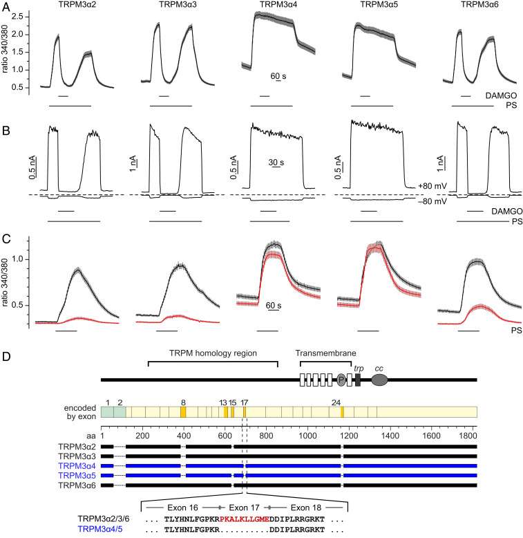
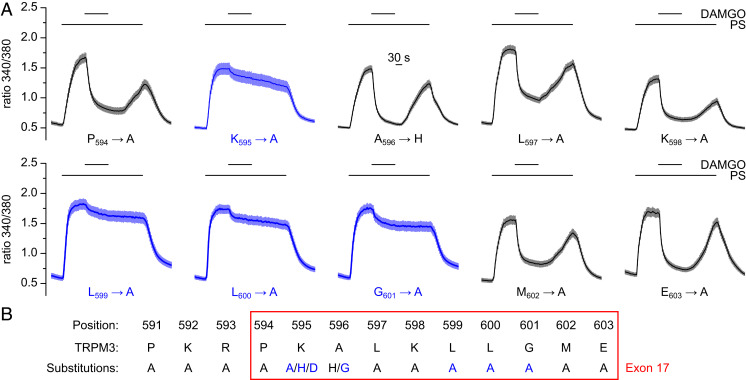
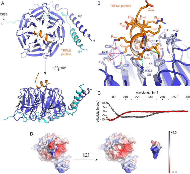
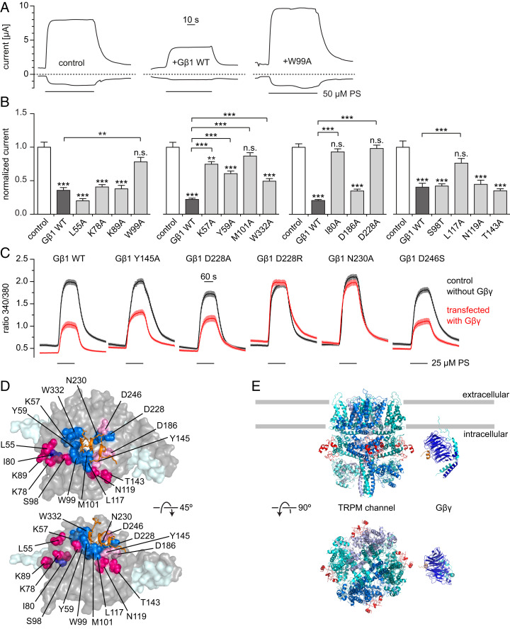
TRPM3 channels play important roles in the detection of noxious heat and in inflammatory thermal hyperalgesia. The activity of these ion channels in somatosensory neurons is tightly regulated by µ-opioid receptors through the signaling of Gβγ proteins, thereby reducing TRPM3-mediated pain. We show here that Gβγ directly binds to a domain of 10 amino acids in TRPM3 and solve a cocrystal structure of this domain together with Gβγ. Using these data and mutational analysis of full-length proteins, we pinpoint three amino acids in TRPM3 and their interacting partners in Gβ1 that are individually necessary for TRPM3 inhibition by Gβγ. The 10-amino-acid Gβγ-interacting domain in TRPM3 is subject to alternative splicing. Its inclusion in or exclusion from TRPM3 channel proteins therefore provides a mechanism for switching on or off the inhibitory action that Gβγ proteins exert on TRPM3 channels.

Keywords: GPCR signaling; TRP channels; alternative splicing; opioid analgesia.


Conflict of interest statement

The authors declare no competing interest.

Figures

Fig. 1.
Fig. 1.
TRPM3 splice variants are differentially susceptible to inhibition by Gβγ. (A) Fura-2 imaging experiments with HEK293 cells cotransfected with the splice variant indicated and µORs show that pregnenolone sulfate (PS)-activated (50 µM PS) TRPM3α4 and TRPM3α5 are not inhibited by µOR activation (3 µM DAMGO), while the other splice variants are profoundly inhibited. (B) Cells similarly transfected were assayed in whole-cell patch-clamp experiments. Each panel depicts a recording of a single representative cell. (C) Fura-2 experiments with cells transfected with Gβγ and a splice variant of TRPM3 (red traces). Black traces show control experiments, in which Gβγ was not transfected. The statistical analysis of the data shown in AC is given in SI Appendix, Fig. S2. (D) Schematic drawing outlining the different splicing events giving rise to the five splice variants investigated (adapted from ref. 33). In the lower half, the amino acid sequences at the junctions of exons 16 and 17 and 17 and 18 are depicted, and amino acids encoded by exon 17 are shown in red. These amino acids are lacking in the splice variants TRPM3α4 and TRPM3α5.
Fig. 2.
Fig. 2.
Individual amino acids encoded by exon 17 are crucial for the GPCR-mediated inhibition of TRPM3 channels. (A) The 10 amino acids encoded by exon 17 were individually mutated (to alanine or histidine, as indicated). After coexpression in HEK293 cells with µORs, the inhibition induced by activation of µORs (3 µM DAMGO) of the pregnenolone sulfate (PS)-induced Ca2+ signals (50 µM PS) was measured. Of the mutants investigated, K595A, L599A, L600A, and G601A showed the strongest defects in the µOR-mediated inhibition. The single-cell analysis of these data is given in SI Appendix, Fig. S6A. Results from further mutations of amino acids in and in the vicinity of exon 17 are reported in SI Appendix, Fig. S6 BE. (B) Summary of the mutations analyzed in A and in SI Appendix, Fig. S6. Blue indicates that the mutation strongly reduced or abolished TRPM3 channel inhibition due to µOR activation.
Fig. 3.
Fig. 3.
Structural analysis of the TRPM3 peptide bound to Gβγ. (A) Overview of the cocrystal structure of Gβγ and a peptide encompassing the TRPM3 exon 17–encoded amino acids in backbone representation with Gβ in blue, Gγ in cyan, and the TRPM3 peptide in orange. (B) A zoom-in to the interaction interface is shown in cartoon and stick representation. For Gβ (blue) only side chains involved in interactions are shown (labeled in blue and underlined for residues undergoing polar interactions and blue and not underlined for residues involved in hydrophobic interactions); for the peptide (orange) all side chains are shown. Polar interactions are indicated with dotted lines for electrostatic interactions (magenta for salt bridges and light magenta for long-range interactions) and hydrogen bonds (green). (C) Secondary structure analysis (using circular dichroism spectroscopy) of the peptide in solution (red dots, alone without interaction partners) shows the signal of a random coil structure. The calculated circular dichroism spectrum of the bound peptide (gray dots) has been included to demonstrate the signal expected from an α-helix. (D) Visualization of surface charges (calculated as “charge-smoothed potential” in PyMOL; red colors indicate negative charges, blue colors positive charges) of the TRPM3 peptide bound to Gβγ (Left) and of both parts displayed separately (Right) demonstrating the complimentary charges on the interacting surfaces. Note that the TRPM3-encoded peptide has been turned by 180° to reveal the charges on the surface that touches Gβ.
Fig. 4.
Fig. 4.
Effects of Gβ mutations on the inhibition of TRPM3. (A) Representative current traces (at −100 and +100 mV) of two-electrode voltage clamp experiments performed in Xenopus oocytes expressing hTRPM3, Gγ2 and wild-type or mutated Gβ1; 50 μM pregnenolone sulfate (PS) was applied as indicated, dashed lines denote zero current. (B) Summary data showing current amplitudes normalized to the average current induced by 50 μM PS in control oocytes without Gβγ coexpression in the same experiment. Mutants tested in the same experiments were grouped into the same panel. Asterisks above columns indicate significant difference from control oocytes without Gβγ expression (*P < 0.05; **P < 0.01; ***P < 0.001; n.s., P ≥ 0.05). (C) Ca2+ imaging experiments in TRPM3-expressing HEK293 cells transiently transfected with Gγ2-IRES-GFP and Myc-tagged Gβ1 (wild-type or mutant, red) or empty vectors as controls (black). When using the mutants D228R and N230A, the strong reduction in PS-induced Ca2+ signals seen with wild-type Gβ was not observed. SI Appendix, Fig. S7 shows single cell responses for the data in AC, as well as expression controls for mutant Gβ1 proteins. (D) Visual summary of the Gβ mutagenesis experiments. Residues that reduced or abolished the inhibition of TRPM3 when mutated are shown as blue spheres, the TRPM3-encoded peptide is drawn as orange sticks. Residues not having an effect on TRPM3 inhibition when mutated are shown as red or pink spheres depending on the apparent distance to the TRPM3 peptide. (E) Cartoon of the proposed model: Gβγ binds to the linker region between MHR3 and MHR4 (red) located on the outer surface of the channel and at a suitable distance from the plasma membrane for binding to membrane-bound Gβγ. Since a 3D structure for TRPM3 is not available, the closely related TRPM7 [PDB ID code 6BWF (49)] is depicted (see SI Appendix, Fig. S8C for additional TRPM structures). The Gβγ structure (blue and cyan) with the TRPM3-encoded peptide (orange) is the 3D structure described in this paper. Gray bars approximately indicate the plasma membrane boundaries.

References

    1. Basbaum A. I., Bautista D. M., Scherrer G., Julius D., Cellular and molecular mechanisms of pain. Cell 139, 267–284 (2009). - PMC - PubMed
    1. Julius D., TRP channels and pain. Annu. Rev. Cell Dev. Biol. 29, 355–384 (2013). - PubMed
    1. Sexton J. E., Vernon J., Wood J. N., TRPs and pain. Handb. Exp. Pharmacol. 223, 873–897 (2014). - PubMed
    1. Vriens J. et al. ., TRPM3 is a nociceptor channel involved in the detection of noxious heat. Neuron 70, 482–494 (2011). - PubMed
    1. Vandewauw I. et al. ., A TRP channel trio mediates acute noxious heat sensing. Nature 555, 662–666 (2018). - PubMed

Publication types

MeSH terms

LinkOut - more resources