. 2020 Oct 29;10(1):18561.
doi: 10.1038/s41598-020-75517-4.

Microglial response to experimental periodontitis in a murine model of Alzheimer's disease

Affiliations

Microglial response to experimental periodontitis in a murine model of Alzheimer's disease

Alpdogan Kantarci et al. Sci Rep. .

Abstract

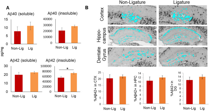
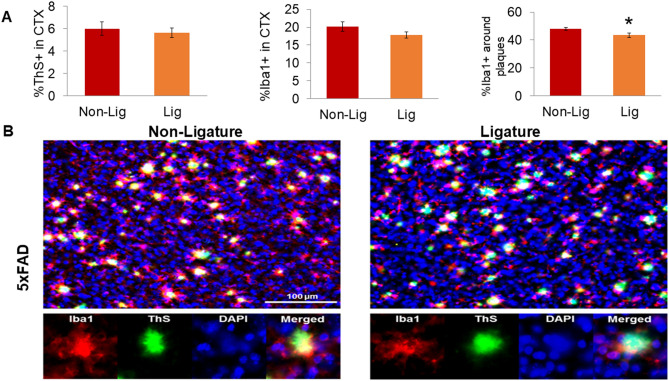
Periodontal disease (PD) has been suggested to be a risk factor for Alzheimer's disease (AD). We tested the impact of ligature-induced PD on 5xFAD mice and WT littermates. At baseline, 5xFAD mice presented significant alveolar bone loss compared to WT mice. After the induction of PD, both WT and 5xFAD mice experienced alveolar bone loss. PD increased the level of Iba1-immunostained microglia in WT mice. In 5xFAD mice, PD increased the level of insoluble Aβ42. The increased level in Iba1 immunostaining that parallels the accumulation of Aβ in 5xFAD mice was not affected by PD except for a decrease in the dentate gyrus. Analysis of double-label fluorescent images showed a decline in Iba1 in the proximity of Aβ plaques in 5xFAD mice with PD compared to those without PD suggesting a PD-induced decrease in plaque-associated microglia (PAM). PD reduced IL-6, MCP-1, GM-CSF, and IFN-γ in brains of WT mice and reduced IL-10 in 5xFAD mice. The data demonstrated that PD increases neuroinflammation in WT mice and disrupts the neuroinflammatory response in 5xFAD mice and suggest that microglia is central to the association between PD and AD.


Conflict of interest statement

The authors declare no competing interests.

Figures

Figure 1
Figure 1
Experimental periodontitis and periodontal changes in a murine model of Alzheimer’s disease. (A) 5xFAD mice at baseline (not exposed to ligatures; Non-Lig) had higher level of bone loss than WT littermates. After induction of experimental periodontitis by the placement of ligatures (Lig), 5xFAD and WT mice both showed significant bone loss while no significant differences were detected between the level of bone loss in ligature-treated 5xFAD mice and ligature-treated WT mice. (B) Macroscopic findings were confirmed with histological measurement of alveolar bone levels in furcation region of the maxillary molar. (C) TRAP stained osteoclastic cells were significantly increased in 5xFAD mice compared to the WT mice at baseline. (*p < 0.05).
Figure 2
Figure 2
Amyloid beta (Aβ) accumulation and topographical distribution in the cortex, hippocampal formation, and dentate gyrus. (A) Soluble and insoluble forms of Aβ in brain extracts from 5xFAD mice with (Lig) or without (Non-Lig) experimental PD. *p < 0.05 compared to non-PD animals. (B) Photomicrographs of Aβ42-immunostained sections containing cortical layers 4–5 (CTX), the hippocampus (HPC), and the dentate gyrus (DG) subregion of the hippocampus, were taken with a 4 × objective from Non-Lig and Lig 5xFAD mouse brain sections. A constant threshold was applied to each image. Analysis of all thresholded Aβ42 particles was performed to obtain the percent area of Aβ42 staining.
Figure 3
Figure 3
Impact of ligature-induced PD on microglia. (A) Photomicrographs of Iba1-immunostained sections from representative mouse brain sections at ×40 magnification (top row). Insets on the first image indicate the magnified (×400) regions-of-interest (ROIs) displayed below as (1) cortical layers 4–5 (CTX), (2) hippocampal CA1, (3) hippocampal fissure (HF), and (4) hippocampal dentate gyrus (DG). (B) Average microglia densitometry was analyzed for each mouse group within the CTX, CA1, HF, and DG ROIs, respectively. Using photomicrographs magnified at ×100, ROIs were outlined, a constant threshold was applied to each image, and analysis of all Iba1+ particles above the threshold was performed to obtain the percent area of Iba1+ staining (microglia densitometry). *p < 0.05, **p < 0.01.
Figure 4
Figure 4
Impact of ligature-induced PD on plaque-associated microglia (PAMs) in 5xFAD mice. Fluorescent photomicrographs containing Iba1+ microglia (red), thioflavin S (ThS) dense-core plaques (green), and DAPI cell nuclei (blue) were taken of cortical layers 4–5 (CTX) from Non-Lig and Lig 5xFAD mice (×20 objective, 2 images/section, 3 sections/animal). Each fluorescent channel was automatically thresholded; an ROI around each ThS+ plaque (including a 6.5 μm buffer-zone) was generated; and channels were analyzed within and without the ROI. (A) Dense-core plaque densitometry (%ThS+), microglia densitometry (%Iba1+), and the percentage of PAMs (%Iba1+ staining within the immediate proximity of ThS+ plaques), *p < 0.05. (B) Representative fluorescent photomicrographs of Non-Lig (left panels) and Lig (right panels) 5xFAD mouse cortex. Scale bar = 100 μm. Bottom row: Iba1, ThS, DAPI, and merged channels from a representative image of a PAM.
Figure 5
Figure 5
Cytokine profile of brain specimens in 5xFAD mice with or without ligature. Higher levels of TNF-α and IL-10, and lower levels of GM-CSF and IFN-γ in the 5xFAD mice brain samples compared to the WT controls were noted prior to the placement of ligatures. There was a significant reduction in IL-6, MCP-1, GM-CSF, and IFN-γ in brains of WT mice and IL-10 in 5xFAD mice after ligature placement. MCP-1 levels in ligated 5xFAD mice were significantly higher than in ligated WT mice. 5xFAD mice showed a higher and unresolved inflammation (TNF-α/IL-10 ratio) compared to WT controls (*p < 0.05, compared to WT animals; #p < 0.05, compared to non-ligature group; ANOVA with posthoc analysis for multiple comparison).

References

    1. Blennow K, de Leon MJ, Zetterberg H. Alzheimer's disease. Lancet. 2006;368:387–403. doi: 10.1016/S0140-6736(06)69113-7. - DOI - PubMed
    1. Akiyama H, et al. Inflammation and Alzheimer's disease. Neurobiol. Aging. 2000;21:383–421. doi: 10.1016/S0197-4580(00)00124-X. - DOI - PMC - PubMed
    1. Kantarci A, et al. Combined administration of resolvin E1 and lipoxin A4 resolves inflammation in a murine model of Alzheimer's disease. Exp. Neurol. 2018;300:111–120. doi: 10.1016/j.expneurol.2017.11.005. - DOI - PubMed
    1. Streit WJ, Xue QS, Tischer J, Bechmann I. Microglial pathology. Acta Neuropathol. Commun. 2014;2:142. doi: 10.1186/s40478-014-0142-6. - DOI - PMC - PubMed
    1. Perry VH, Nicoll JA, Holmes C. Microglia in neurodegenerative disease. Nat. Rev. Neurol. 2010;6:193–201. doi: 10.1038/nrneurol.2010.17. - DOI - PubMed

Publication types

MeSH terms