. 2020 Oct 29;10(1):18620.
doi: 10.1038/s41598-020-75676-4.

Augmented reality improves procedural efficiency and reduces radiation dose for CT-guided lesion targeting: a phantom study using HoloLens 2

Affiliations

Augmented reality improves procedural efficiency and reduces radiation dose for CT-guided lesion targeting: a phantom study using HoloLens 2

Brian J Park et al. Sci Rep. .

Abstract

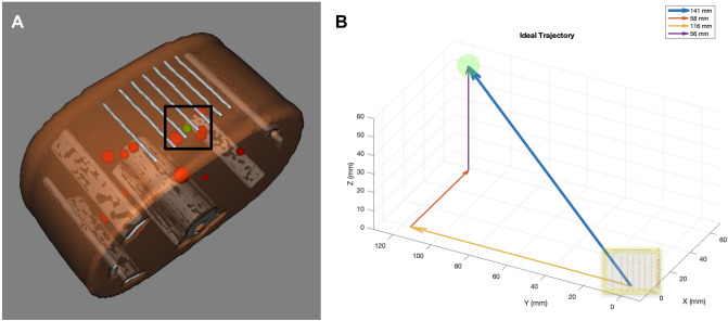
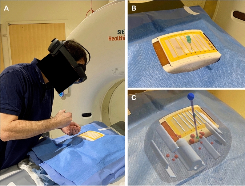
Out-of-plane lesions pose challenges for CT-guided interventions. Augmented reality (AR) headsets are capable to provide holographic 3D guidance to assist CT-guided targeting. A prospective trial was performed assessing CT-guided lesion targeting on an abdominal phantom with and without AR guidance using HoloLens 2. Eight operators performed a cumulative total of 86 needle passes. Total needle redirections, radiation dose, procedure time, and puncture rates of nontargeted lesions were compared with and without AR. Mean number of needle passes to reach the target reduced from 7.4 passes without AR to 3.4 passes with AR (p = 0.011). Mean CT dose index decreased from 28.7 mGy without AR to 16.9 mGy with AR (p = 0.009). Mean procedure time reduced from 8.93 min without AR to 4.42 min with AR (p = 0.027). Puncture rate of a nontargeted lesion decreased from 11.9% without AR (7/59 passes) to 0% with AR (0/27 passes). First needle passes were closer to the ideal target trajectory with AR versus without AR (4.6° vs 8.0° offset, respectively, p = 0.018). AR reduced variability and elevated the performance of all operators to the same level irrespective of prior clinical experience. AR guidance can provide significant improvements in procedural efficiency and radiation dose savings for targeting out-of-plane lesions.


Conflict of interest statement

The authors declare no competing interests.

Figures

Figure 1
Figure 1
CT phantom abdominal biopsy phantom. (A) CT grid is applied to the surface of the model. Phantom contains multiple targets of various sizes. (B) CT image of model. Selected target measures 11 mm in diameter.
Figure 2
Figure 2
Three-dimensional surface-rendered model of phantom. (A) Lines from the CT grid can be seen along the anterior surface. Target lesion is specified in green. All other nontargeted lesions are specified in red. (B) Wireframe view of model which contains 58,498 polygons with a total file size of only 1.6 MB.
Figure 3
Figure 3
Trajectory to targeted lesion from specified skin entry site. (A) Down-the-barrel look at trajectory to targeted lesion (green) from skin entry site at the inferior aspect between labeled gridlines 3 and 4 (black box). Several nontargeted lesions (red) can be seen in close proximity to the trajectory. (B) Vector of ideal trajectory based on preoperative CT scan from specified skin entry site. Total trajectory distance of 14.1 cm from skin with 23.4° angle relative to the z-plane (5.8 cm lateral, 11.6 cm deep, and 5.6 cm cranial component). Target and CT grid are not drawn to scale.
Figure 4
Figure 4
Augmented reality (AR)-assisted navigation using HoloLens 2. (A) Participant inserts the needle while wearing HoloLens 2. (B) View of needle insertion without AR. (C) View of needle insertion through HoloLens 2 with three-dimensional model and virtual needle guide projected onto the phantom. Registration is visually confirmed with the actual CT gridlines aligned with the virtual gridlines. The needle is seen aligned with the virtual guide (purple line) displaying the ideal trajectory to the target lesion (green ball). Note that this two-dimensional captured image does not fully represent the three-dimensional stereoscopic view seen with HoloLens 2.
Figure 5
Figure 5
Flowchart of study design. Order of interventions with and without augmented reality-assisted navigation were randomized to limit order bias.
Figure 6
Figure 6
Diagram demonstrating calculations in two dimensions for illustrative purposes only. Actual calculations were performed in three dimensions based on voxel locations. Blue solid arrow represents distance of needle tip from skin entry site. Red solid arrow represents remaining distance to center of target. Yellow dotted arrow represents ideal trajectory from skin entry site to center of target. Angle offsets were calculated between the needle trajectory (blue solid arrow) relative to the ideal trajectory (yellow dotted arrow).

References

    1. Das M, et al. Augmented reality visualization for CT-guided interventions: system description, feasibility, and initial evaluation in an abdominal phantom. Radiology. 2006;240:230–235. doi: 10.1148/radiol.2401040018. - DOI - PubMed
    1. Uppot RN, et al. Implementing virtual and augmented reality tools for radiology education and training, communication, and clinical care. Radiology. 2019;291:570–580. doi: 10.1148/radiol.2019182210. - DOI - PubMed
    1. Park BJ, et al. Augmented and mixed reality: technologies for enhancing the future of IR. J. Vasc. Interv. Radiol. 2020;31:1074–1082. doi: 10.1016/j.jvir.2019.09.020. - DOI - PMC - PubMed
    1. Maybody M, Stevenson C, Solomon SB. Overview of navigation systems in image-guided interventions. Tech. Vasc. Interv. Radiol. 2013;16:136–143. doi: 10.1053/j.tvir.2013.02.008. - DOI - PubMed
    1. Durand P, et al. Computer assisted electromagnetic navigation improves accuracy in computed tomography guided interventions: a prospective randomized clinical trial. PLoS ONE. 2017;12:e0173751. doi: 10.1371/journal.pone.0173751. - DOI - PMC - PubMed

Publication types