. 2020 Oct 29;10(1):18636.
doi: 10.1038/s41598-020-75725-y.

Contralateral spreading of substances following intratympanic nanoparticle-conjugated gentamicin injection in a rat model

Affiliations

Contralateral spreading of substances following intratympanic nanoparticle-conjugated gentamicin injection in a rat model

Sang-Yeon Lee et al. Sci Rep. .

Abstract

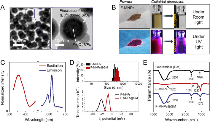
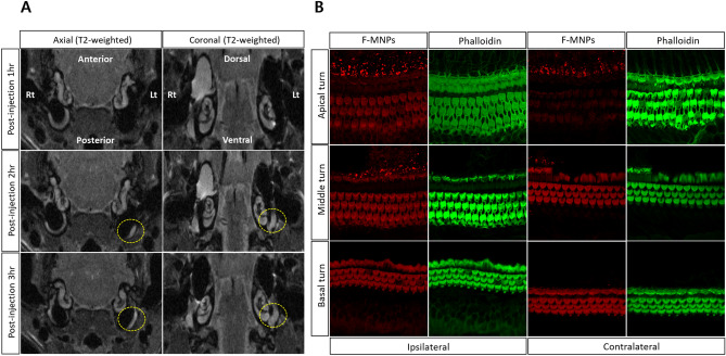
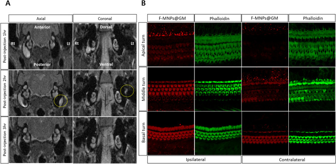
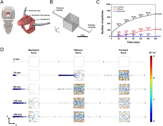
This study was performed to investigate the Eustachian tube as a potential route for contralateral spreading following intratympanic nanoparticle (NP)-conjugated gentamicin injection in a rat model. Sprague-Dawley rats were divided into three groups and substances were injected in the right ear: group 1 (fluorescent magnetic nanoparticles [F-MNPs], n = 4), group 2 (F-MNP-conjugated gentamicin [F-MNP@GM], n = 2), and control group (no injections, n = 2). T2-weighted sequences corresponding to the regions of interest at 1, 2, and 3 h after intratympanic injection were evaluated, along with immunostaining fluorescence of both side cochlea. The heterogeneous signal intensity of F-MNPs and F-MNP@GM on T2-weighted images, observed in the ipsilateral tympanum, was also detected in the contralateral tympanum in 4 out of 6 rats, recapitulating fluorescent nanoparticles in the contralateral cochlear hair cells. Computational simulations demonstrate the contralateral spreading of particles by gravity force following intratympanic injection in a rat model. The diffusion rate of the contralateral spreading relies on the sizes and surface charges of particles. Collectively, the Eustachian tube could be a route for contralateral spreading following intratympanic injection. Caution should be taken when using the contralateral ear as a control study investigating inner-ear drug delivery through the transtympanic approach.


Conflict of interest statement

One of the authors (S.I.K) is somehow related to the AMO LIFE SCIENCE Co. However, the author(s) declare no competing interests.

Figures

Figure 1
Figure 1
Characterization of fluorescent magnetic nanoparticles (F-MNPs) and F-MNP-conjugated gentamicin (F-MNP@GM). (A) Transmission electron microscopy (TEM) images of F-MNPs at different magnifications. (B) Photographs of the particle powder and dispersion under visible and UV light. (C) Excitation and emission spectra of F-MNPs. (D) Hydrodynamic diameter, zeta potential, and (E) Fourier transform infrared (FT-IR) spectrum of F-MNPs during the process of drug loading.
Figure 2
Figure 2
(A) Temporal visualization of intratympanically injected fluorescent magnetic nanoparticles (F-MNPs) that spread to the contralateral tympanum. Specifically, the yellow dotted circle represents the signal intensity relevant F-MNPs in the contralateral tympanum at the same sequence representing the axial and coronal scans, respectively. (B) Immunostaining of cochlear hair cells for each turn of ipsilateral and contralateral cochlear hair cells after intratympanic injection of F-MNPs. This figure represents rat number 2 in group 1.
Figure 3
Figure 3
(A) Temporal visualization of intratympanically injected fluorescent magnetic nanoparticles (F-MNPs)-conjugated gentamicin (F-MNP@GM) spreading to the contralateral tympanum. Specifically, the yellow dotted circle represents the signal intensity relevant F-MNP@GM in the contralateral tympanum at the same sequence representing the axial and coronal scans, respectively. (B) Immunostaining of cochlear hair cells for each turn of ipsilateral and contralateral cochlear hair cells after intratympanic injection of F-MNP@GM. No F-MNP@GM-related fluorescence was depicted in the contralateral apical turn of the cochlea. This figure represents rat number 1 in group 2.
Figure 4
Figure 4
The simulation results of particle diffusion in the Eustachian tube. (A) The scheme of Eustachian tube anatomy of the rat. (B) Eustachian tube model for simulation geometry. (C) The number of particles arrived at the opposite side of the Eustachian tube by the time. (D) The simulation snapshots of particle diffusion in the Eustachian tube depending on time. For more clarify, the size of nanoparticles is denoted as a magnified scale. The color legend shows the position of the particle (y2 + z2) from the Eustachian tube axis (x-axis).
Figure 5
Figure 5
Hypothetical explanation of the contralateral spreading via the eustachian tube following the ipsilateral transtympanic injection of nanoparticle conjugated gentamycin.

References

    1. Sajjadi H, Paparella MM. Meniere's disease. Lancet. 2008;372:406–414. doi: 10.1016/S0140-6736(08)61161-7. - DOI - PubMed
    1. Yardley L, Dibb B, Osborne G. Factors associated with quality of life in Meniere's disease. Clin. Otolaryngol. Allied Sci. 2003;28:436–441. doi: 10.1046/j.1365-2273.2003.00740.x. - DOI - PubMed
    1. Cohen-Kerem R, et al. Intratympanic gentamicin for Meniere's disease: A meta-analysis. Laryngoscope. 2004;114:2085–2091. doi: 10.1097/01.mlg.0000149439.43478.24. - DOI - PubMed
    1. Rah YC, Han JJ, Park J, Choi BY, Koo JW. Management of intractable M énière's disease after intratympanic injection of Gentamicin. Laryngoscope. 2015;125:972–978. doi: 10.1002/lary.25009. - DOI - PubMed
    1. Salt A, et al. Perilymph pharmacokinetics of locally-applied gentamicin in the guinea pig. Hear. Res. 2016;342:101–111. doi: 10.1016/j.heares.2016.10.003. - DOI - PMC - PubMed

Publication types