. 2020 Oct 29;10(1):18693.
doi: 10.1038/s41598-020-75747-6.

Computational modelling of patient specific spring assisted lambdoid craniosynostosis correction

Affiliations

Computational modelling of patient specific spring assisted lambdoid craniosynostosis correction

Selim Bozkurt et al. Sci Rep. .

Abstract

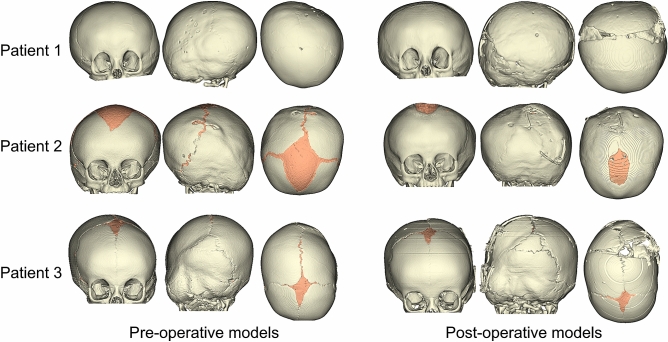
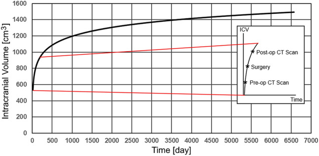
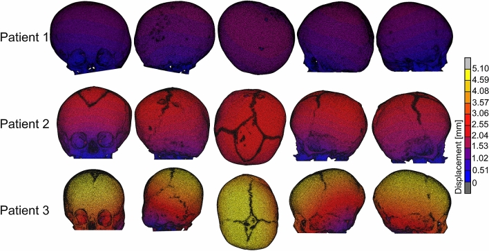
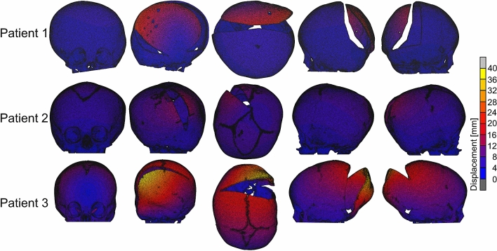
Lambdoid craniosynostosis (LC) is a rare non-syndromic craniosynostosis characterised by fusion of the lambdoid sutures at the back of the head. Surgical correction including the spring assisted cranioplasty is the only option to correct the asymmetry at the skull in LC. However, the aesthetic outcome from spring assisted cranioplasty may remain suboptimal. The aim of this study is to develop a parametric finite element (FE) model of the LC skulls that could be used in the future to optimise spring surgery. The skull geometries from three different LC patients who underwent spring correction were reconstructed from the pre-operative computed tomography (CT) in Simpleware ScanIP. Initially, the skull growth between the pre-operative CT imaging and surgical intervention was simulated using MSC Marc. The osteotomies and spring implantation were performed to simulate the skull expansion due to the spring forces and skull growth between surgery and post-operative CT imaging in MSC Marc. Surface deviation between the FE models and post-operative skull models reconstructed from CT images changed between ± 5 mm over the skull geometries. Replicating spring assisted cranioplasty in LC patients allow to tune the parameters for surgical planning, which may help to improve outcomes in LC surgeries in the future.


Conflict of interest statement

The authors declare no competing interests.

Figures

Figure 1
Figure 1
Patient-specific pre and post-operative skull models reconstructed from the CT images.
Figure 2
Figure 2
Intracranial volume (ICV) simulated by the model in Eq. (1) and region of interest on the growth curve with representative pre-operative, surgical intervention and post-operative times.
Figure 3
Figure 3
The FE models simulating spring assisted cranial expansion with osteotomies.
Figure 4
Figure 4
Shapes of the intracranial cavities in the patients’ skulls.
Figure 5
Figure 5
Displacement maps for the FE models simulating the skull growth between the pre-operative CT scan and surgical intervention.
Figure 6
Figure 6
Surface deviation between the FE models and post-operative skull models reconstructed from CT images.
Figure 7
Figure 7
Comparison between the cross-sections of the FE models simulating spring assisted cranioplasty (orange) and post-operative skull growth and post-operative skull models reconstructed from CT images (black).
Figure 8
Figure 8
Displacement maps for the skull models for the spring assisted cranioplasty and post-operative skull growth.

References

    1. David DJ, Menard RM. Occipital plagiocephaly. Br. J. Plast. Surg. 2000;53:367–377. doi: 10.1054/bjps.2000.3329. - DOI - PubMed
    1. Boulet SL, Rasmussen SA, Honein MA. A population-based study of craniosynostosis in metropolitan Atlanta, 1989–2003. Am. J. Med. Genet. A. 2008;146A:984–991. doi: 10.1002/ajmg.a.32208. - DOI - PubMed
    1. Borad V, et al. Isolated lambdoid craniosynostosis. J. Craniofac. Surg. 2019;30:2390–2392. doi: 10.1097/SCS.0000000000006058. - DOI - PubMed
    1. Rhodes JL, Tye GW, Fearon JA. Craniosynostosis of the lambdoid suture. Semin. Plast. Surg. 2014;28:138–143. doi: 10.1055/s-0034-1384809. - DOI - PMC - PubMed
    1. Ghizoni E, et al. Diagnosis of infant synostotic and nonsynostotic cranial deformities: a review for pediatricians. Rev. Paul. Pediatr. 2016;34:495–502. - PMC - PubMed

Publication types