. 2020 Oct 23:15:8175-8200.
doi: 10.2147/IJN.S269630. eCollection 2020.

Thermosensitive Betulinic Acid-Loaded Magnetoliposomes: A Promising Antitumor Potential for Highly Aggressive Human Breast Adenocarcinoma Cells Under Hyperthermic Conditions

Affiliations

Thermosensitive Betulinic Acid-Loaded Magnetoliposomes: A Promising Antitumor Potential for Highly Aggressive Human Breast Adenocarcinoma Cells Under Hyperthermic Conditions

Claudia Geanina Farcas et al. Int J Nanomedicine. .

Abstract

Purpose: Breast cancer presents one of the highest rates of prevalence around the world. Despite this, the current breast cancer therapy is characterized by significant side effects and high risk of recurrence. The present work aimed to develop a new therapeutic strategy that may improve the current breast cancer therapy by developing a heat-sensitive liposomal nano-platform suitable to incorporate both anti-tumor betulinic acid (BA) compound and magnetic iron nanoparticles (MIONPs), in order to address both remote drug release and hyperthermia-inducing features. To address the above-mentioned biomedical purposes, the nanocarrier must possess specific features such as specific phase transition temperature, diameter below 200 nm, superparamagnetic properties and heating capacity. Moreover, the anti-tumor activity of the developed nanocarrier should significantly affect human breast adenocarcinoma cells.

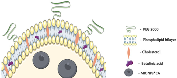
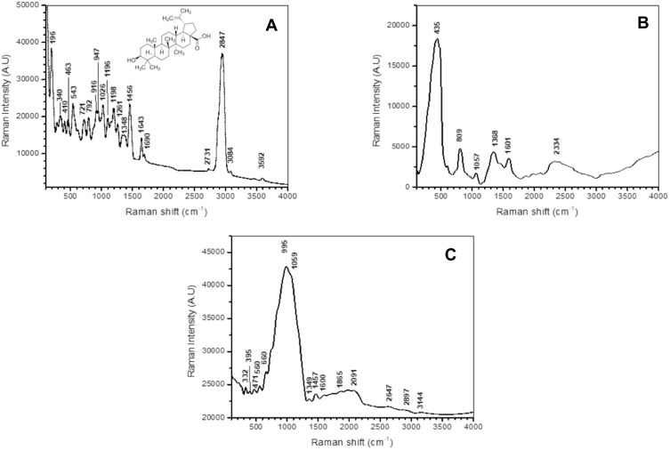
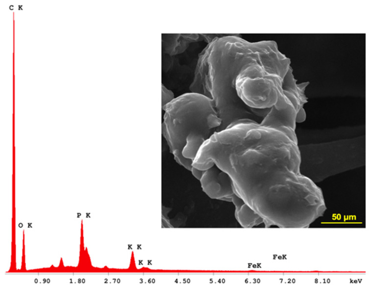
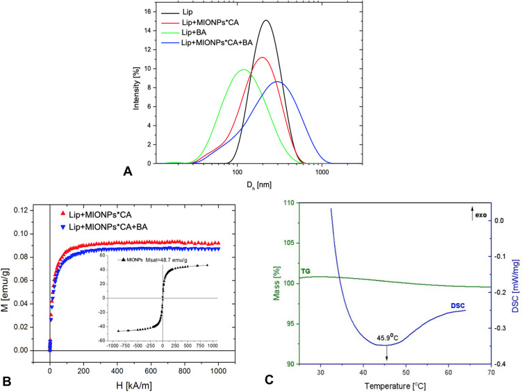
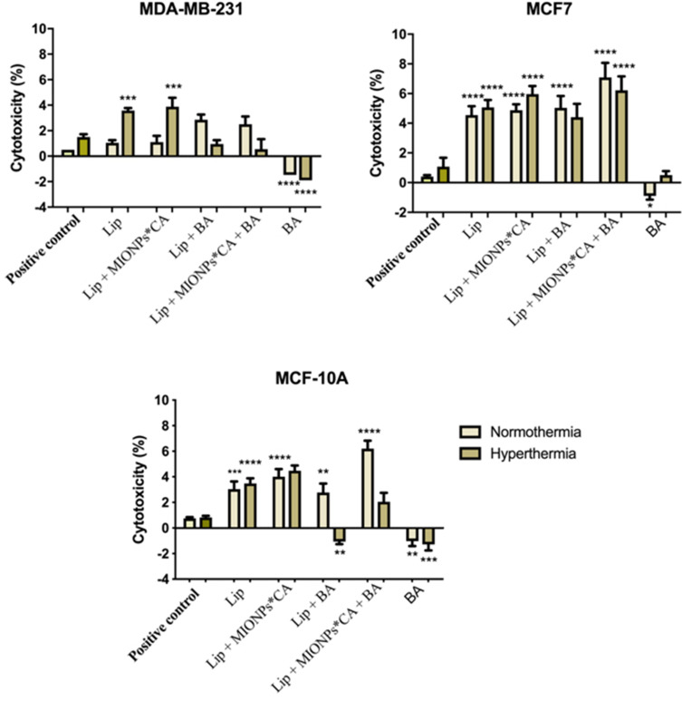
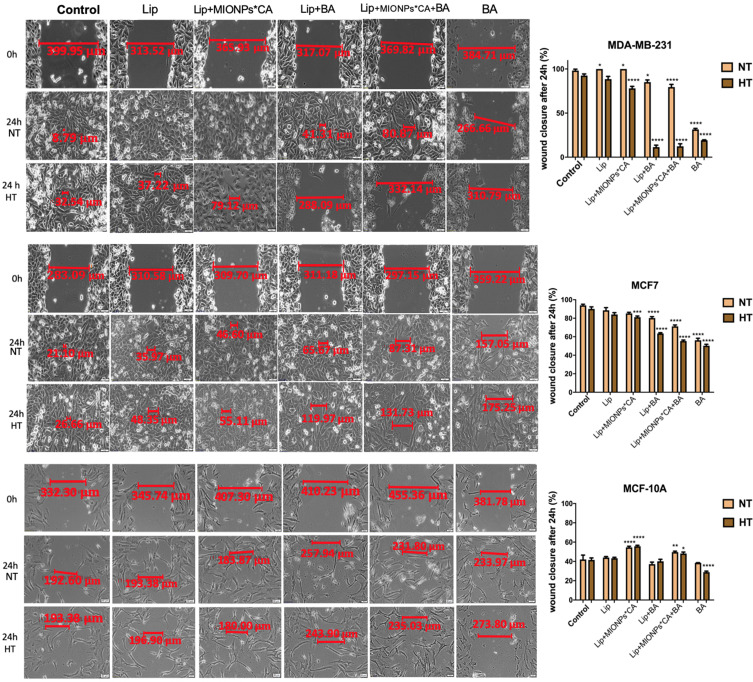
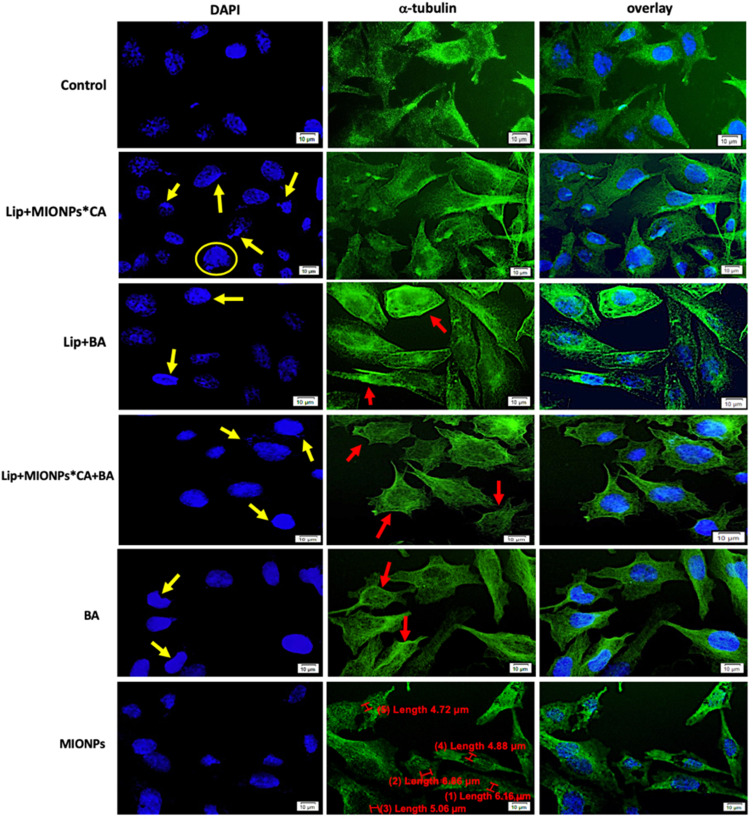
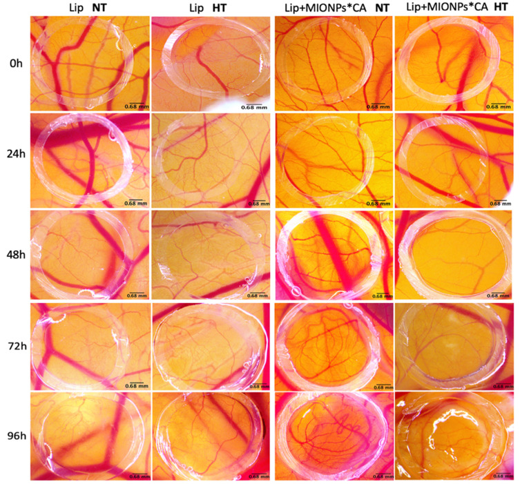
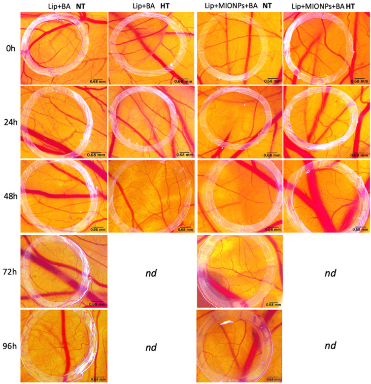
Methods: BA-loaded magnetoliposomes and corresponding controls (BA-free liposomes and liposomes containing no magnetic payload) were obtained through the thin-layer hydration method. The quality and stability of the multifunctional platforms were physico-chemically analysed by the means of RAMAN, scanning electron microscopy-EDAX, dynamic light scattering, zeta potential and DSC analysis. Besides this, the magnetic characterization of magnetoliposomes was performed in terms of superparamagnetic behaviour and heating capacity. The biological profile of the platforms and controls was screened through multiple in vitro methods, such as MTT, LDH and scratch assays, together with immunofluorescence staining. In addition, CAM assay was performed in order to assess a possible anti-angiogenic activity induced by the test samples.

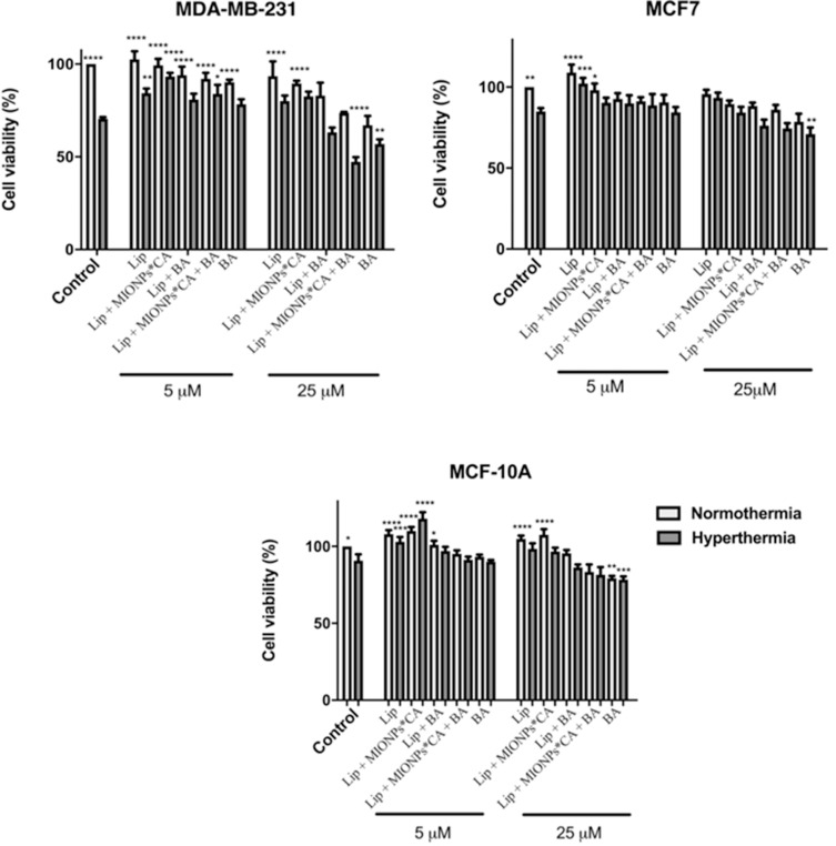
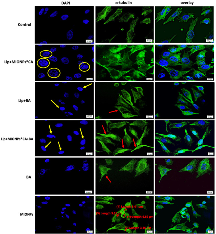
Results: The physico-chemical analysis revealed that BA-loaded magnetoliposomes present suitable characteristics for the purpose of this study, showing biocompatible phase transition temperature, a diameter of 198 nm, superparamagnetic features and heating capacity. In vitro results showed that hyperthermia induces enhanced anti-tumor activity when breast adenocarcinoma MDA-MB-231 cells were exposed to BA-loaded magnetoliposomes, while a low cytotoxic rate was exhibited by the non-tumorigenic breast epithelial MCF 10A cells. Moreover, the in ovo angiogenesis assay endorsed the efficacy of this multifunctional platform as a good strategy for breast cancer therapy, under hyperthermal conditions. Regarding the possible mechanism of action of this multifunctional nano-platform, the immunocytochemistry of the MCF7 and MDA-MB-231 breast carcinoma cells revealed a microtubule assembly modulatory activity, under hyperthermal conditions.

Conclusion: Collectively, these findings indicate that BA-loaded magnetoliposomes, under hyperthermal conditions, might serve as a promising strategy for breast adenocarcinoma treatment.

Keywords: betulinic acid; breast adenocarcinoma; hyperthermia; magnetoliposomes.


Conflict of interest statement

The authors report no conflicts of interest in this work.

Figures

Figure 1
Figure 1
Graphical representation of a BA-loaded magnetoliposome. MIONPs*CA are incorporated in the aqueous core, while BA is entrapped in the lipid bilayer of the liposome. (Image realized by applying Servier Medical ART illustration: http://smart.servier.com).
Figure 2
Figure 2
(A) Molecular structure and Micro-Raman spectra of betulinic acid (BA); (B) Micro-Raman spectra of magnetic iron oxide nanoparticles (MIONPs); (C) Micro-Raman spectra of BA-loaded magnetoliposomes.
Figure 3
Figure 3
SEM-EDAX analysis of BA-loaded magnetoliposomes.
Figure 4
Figure 4
Graphical representation of: (A) intensity distribution of particles size of the liposomal samples; (B) magnetization curves of magnetoliposomes and control (MIONPs); (C) TG/DSC curves of BA-loaded magnetoliposome.
Figure 5
Figure 5
Viability percentages of breast adenocarcinoma (MDA-MB-231, MCF7) cells and breast epithelial (MCF 10A) cells after exposure to liposomal structures at concentrations of 5 and 25 µM. The MTT assessment was performed 24h post-stimulation, under normothermic (37 °C) and hyperthermic (43 °C) conditions. The cell viability percentage was normalized to control cells (no stimulation, under normothermic conditions). One-way ANOVA analysis was applied to determine the statistical differences followed by Tukey’s multiple comparisons test vs hyperthermia control (*p < 0.05; **p < 0.01; ***p < 0.001, ****p < 0.0001). The data represent the mean values ± SD of three independent experiments (n=3).
Figure 6
Figure 6
Cytotoxicity percentages of breast adenocarcinoma (MDA-MB-231, MCF7) cells and breast epithelial (MCF 10A) cells after exposure to liposomal structures at concentration of 25 µM. The LDH assessment was performed 24 h post-stimulation under normothermic (37 °C) and hyperthermic (43 °C) conditions. One-way ANOVA analysis was applied to determine the statistical differences followed by Tukey’s multiple comparisons test vs hyperthermia control (*p < 0.05; **p < 0.01; ***p < 0.001, ****p < 0.0001). The data represent the mean values ± SD of three independent experiments (n=3).
Figure 7
Figure 7
Representative images of the migratory capacity of the breast adenocarcinoma (MDA-MB-231, MCF7) cells and breast epithelial (MCF 10A) cells after treatment with test compounds at concentration of 25 µM. The results were expressed as percentage of wound closure after 24 h (normothermia/hyperthermia) compared to the initial surface; NT – normothermia (37 °C), HT – hyperthermia (43 °C). The cells were visualized by light microscopy, at magnification 20x. Scale bars represent 50 µm. One-way ANOVA analysis was applied to determine the statistical differences followed by Tukey’s multiple comparisons test vs hyperthermia control (*p < 0.05; **p < 0.01; ***p < 0.001, ****p < 0.0001). The data represent the mean values ± SD of three independent experiments (n=3).
Figure 8
Figure 8
MDA-MB-231 cells visualized by fluorescence microscopy, after 24 h treatment with test compounds at concentration of 25 µM, under hyperthermal conditions. Nuclear and microtubules staining were expressed separately (DAPI and α-tubulin, respectively) and also combined (overlay). Yellow arrows marked the typical morphological changes for apoptosis induction: chromatin condensation, boundary alterations, and nuclear fragmentation, while enucleation process is highlighted by the yellow circle. The abnormal filamentous organization of microtubule (MT) network was indicated by the red arrows. Three independent experiments were performed for each sample (n=3).
Figure 9
Figure 9
MCF7 cells visualized by fluorescence microscopy, after 24 h treatment with test compounds at concentration of 25 µM under hyperthermal conditions. Nuclear and microtubules staining were expressed separately (DAPI and α-tubulin, respectively) and also combined (overlay). Yellow arrows marked the typical morphological changes for apoptosis induction: chromatin condensation, boundary alterations, and nuclear fragmentation, while enucleation process is highlighted by the yellow circle. The abnormal filamentous organization of microtubule (MT) network was indicated by the red arrows. Three independent experiments were performed for each sample (n=3).
Figure 10
Figure 10
Stereomicroscopic in ovo images of the vascularized areas treated with blank liposomal samples (Lip and Lip+MIONPs*CA) at concentration of 25 µM, under normothermic – NT (37 °C) and hyperthermic pre-treatment – HT (46 °C). Three independent experiments were performed for each sample (n=3).
Figure 11
Figure 11
Stereomicroscopic in vivo images of the vascularized areas treated with liposomal samples containing BA (Lip+BA and Lip+MIONPs*CA+BA) at concentration of 25 µM, under normothermic - NT (37 °C) and hyperthermic pre-treatment - HT (46 °C). The specimens with hyperthermic pre-treatment died after 48h, so they were not determined (nd) further. Three independent experiments were performed for each sample (n=3).

Similar articles

Cited by

References

    1. Temian DC, Pop LA, Irimie AI, Berindan-Neagoe I. The epigenetics of triple-negative and basal-like breast cancer: current knowledge. J Breast Cancer. 2018;21(3):233–243. doi:10.4048/jbc.2018.21.e41 - DOI - PMC - PubMed
    1. Braicu C, Chiorean R, Irimie A, et al. Novel insight into triple-negative breast cancers, the emerging role of angiogenesis, and antiangiogenic therapy. Expert Rev Mol Med. 2016;18:e18. doi:10.1017/erm.2016.17 - DOI - PubMed
    1. WHO. Breast cancer: prevention and control; 2020. Available from: https://www.who.int/cancer/detection/breastcancer/en/. Accessed October8, 2020.
    1. Bandyopadhyay S, Bluth MH, Ali-Fehmi R. Breast carcinoma: updates in molecular profiling 2018. Clin Lab Med. 2018;38(2):401–420. doi:10.1016/j.cll.2018.02.006 - DOI - PubMed
    1. Hurvitz S, Mead M. Triple-negative breast cancer: advancements in characterization and treatment approach. Curr Opin Obstet Gynecol. 2016;28(1):59–69. doi:10.1097/GCO.0000000000000239 - DOI - PubMed

MeSH terms