Tissue Clearing and Expansion Methods for Imaging Brain Pathology in Neurodegeneration: From Circuits to Synapses and Beyond
- PMID: 33122983
- PMCID: PMC7571329
- DOI: 10.3389/fnins.2020.00914
Tissue Clearing and Expansion Methods for Imaging Brain Pathology in Neurodegeneration: From Circuits to Synapses and Beyond
Abstract
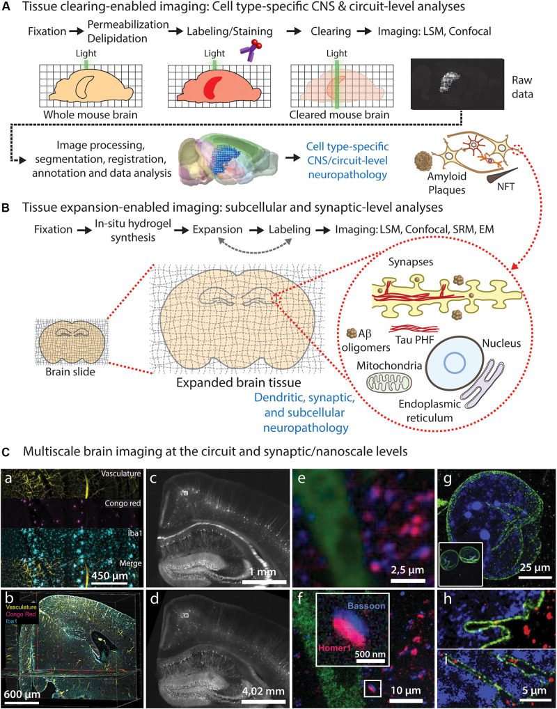
Studying the structural alterations occurring during diseases of the nervous system requires imaging heterogeneous cell populations at the circuit, cellular and subcellular levels. Recent advancements in brain tissue clearing and expansion methods allow unprecedented detailed imaging of the nervous system through its entire scale, from circuits to synapses, including neurovascular and brain lymphatics elements. Here, we review the state-of-the-art of brain tissue clearing and expansion methods, mentioning their main advantages and limitations, and suggest their parallel implementation for circuits-to-synapses brain imaging using conventional (diffraction-limited) light microscopy -such as confocal, two-photon and light-sheet microscopy- to interrogate the cellular and molecular basis of neurodegenerative diseases. We discuss recent studies in which clearing and expansion methods have been successfully applied to study neuropathological processes in mouse models and postmortem human brain tissue. Volumetric imaging of cleared intact mouse brains and large human brain samples has allowed unbiased assessment of neuropathological hallmarks. In contrast, nanoscale imaging of expanded cells and brain tissue has been used to study the effect of protein aggregates on specific subcellular structures. Therefore, these approaches can be readily applied to study a wide range of brain processes and pathological mechanisms with cellular and subcellular resolution in a time- and cost-efficient manner. We consider that a broader implementation of these technologies is necessary to reveal the full landscape of cellular and molecular mechanisms underlying neurodegenerative diseases.
Keywords: expansion microscopy; neurodegeneration; neuropathology; super-resolution microscopy; tissue clearing.
Copyright © 2020 Parra-Damas and Saura.
Figures

Similar articles
-
Expansion Light Sheet Microscopy Resolves Subcellular Structures in Large Portions of the Songbird Brain.Front Neuroanat. 2019 Jan 31;13:2. doi: 10.3389/fnana.2019.00002. eCollection 2019. Front Neuroanat. 2019. PMID: 30766480 Free PMC article.
-
Whole-Brain Single-Cell Imaging and Analysis of Intact Neonatal Mouse Brains Using MRI, Tissue Clearing, and Light-Sheet Microscopy.J Vis Exp. 2022 Aug 1;(186):10.3791/64096. doi: 10.3791/64096. J Vis Exp. 2022. PMID: 35969091 Free PMC article.
-
Light-sheet fluorescence expansion microscopy: fast mapping of neural circuits at super resolution.Neurophotonics. 2019 Jan;6(1):015005. doi: 10.1117/1.NPh.6.1.015005. Epub 2019 Feb 8. Neurophotonics. 2019. PMID: 30796881 Free PMC article.
-
Current Status of Tissue Clearing and the Path Forward in Neuroscience.ACS Chem Neurosci. 2021 Jan 6;12(1):5-29. doi: 10.1021/acschemneuro.0c00563. Epub 2020 Dec 16. ACS Chem Neurosci. 2021. PMID: 33326739 Review.
-
[Getting an insight into the brain - new optical clearing techniques and imaging using light-sheet microscope].Postepy Biochem. 2017;63(1):8-15. Postepy Biochem. 2017. PMID: 28409571 Review. Polish.
Cited by
-
Development of Planar Illumination Strategies for Solving Mysteries in the Sub-Cellular Realm.Int J Mol Sci. 2022 Jan 31;23(3):1643. doi: 10.3390/ijms23031643. Int J Mol Sci. 2022. PMID: 35163562 Free PMC article. Review.
-
Network Neuroscience Untethered: Brain-Wide Immediate Early Gene Expression for the Analysis of Functional Connectivity in Freely Behaving Animals.Biology (Basel). 2022 Dec 24;12(1):34. doi: 10.3390/biology12010034. Biology (Basel). 2022. PMID: 36671727 Free PMC article. Review.
-
Towards organism-level systems biology by next-generation genetics and whole-organ cell profiling.Biophys Rev. 2021 Nov 18;13(6):1113-1126. doi: 10.1007/s12551-021-00859-w. eCollection 2021 Dec. Biophys Rev. 2021. PMID: 35059031 Free PMC article. Review.
-
Ultraviolet photoacoustic microscopy with tissue clearing for high-contrast histological imaging.Photoacoustics. 2021 Nov 3;25:100313. doi: 10.1016/j.pacs.2021.100313. eCollection 2022 Mar. Photoacoustics. 2021. PMID: 34804794 Free PMC article.
-
Comparison of Different Tissue Clearing Methods for Three-Dimensional Reconstruction of Human Brain Cellular Anatomy Using Advanced Imaging Techniques.Front Neuroanat. 2021 Nov 11;15:752234. doi: 10.3389/fnana.2021.752234. eCollection 2021. Front Neuroanat. 2021. PMID: 34867215 Free PMC article.
References
Publication types
LinkOut - more resources
Full Text Sources
Other Literature Sources