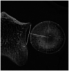
Positioning of longest axis of the radial head in neutral forearm rotation
- PMID: 33123224
- PMCID: PMC7545526
- DOI: 10.1177/1758573219831285
Positioning of longest axis of the radial head in neutral forearm rotation
Abstract
Introduction: The radial head has an ellipsoid shape so that a longest and a shortest axis can be defined. The aim of this study is to evaluate the position of the longest axis of the radial head (LARH) in relation to proximal radioulnar joint (PRUJ) and to the forearm in neutral position using 3D computed tomography (CT).
Materials and methods: 3D CT reconstructions of the distal humerus, the radius and the ulna of 27 healthy volunteers (average age 27.65 ± 9.25; 24 males, 3 females) were created. First an evaluation of the elliptic form of the radial head and the location of its longest axis was performed. Next, three planes were defined: the PRUJ plane, the forearm plane and a neutral plane. Based on the angle between the forearm plane and the neutral plane, the rotation of the scanned forearm was measured. Taking this rotation into account, the position of the LARH compared to PRUJ plane and forearm plane in neutral position is recalculated.
Results: The shape of the radial head is determined to be non-circular based on this study population (p < .001). In neutral position, the angle between the LARH and the forearm plane is 5.28° (SD: 15.09) and between the LARH and the PRUJ is 33.46° (SD: 13.91).
Conclusions: The position of the LARH is found to be approximately perpendicular to the forearm plane when the forearm is in neutral position and perpendicular to the PRUJ plane when the forearm is on average in 30° of pronation.
Keywords: 3D reconstructed; CT; Radial head; neutral rotation; non-circular; proximal radioulnar joint.
© 2019 The British Elbow & Shoulder Society.
Figures


Similar articles
-
The role of radial head morphology in proximal radioulnar joint congruency during forearm rotation.J Exp Orthop. 2024 Oct 21;11(4):e70059. doi: 10.1002/jeo2.70059. eCollection 2024 Oct. J Exp Orthop. 2024. PMID: 39435300 Free PMC article.
-
Influence of forearm rotation on proximal radioulnar joint congruency and translational motion using computed tomography and computer-aided design technologies.J Hand Surg Am. 2011 May;36(5):811-5. doi: 10.1016/j.jhsa.2011.01.043. J Hand Surg Am. 2011. PMID: 21527137
-
Where Is the Ulnar Styloid Process? Identification of the Absolute Location of the Ulnar Styloid Process Based on CT and Verification of Neutral Forearm Rotation on Lateral Radiographs of the Wrist.Clin Orthop Surg. 2018 Mar;10(1):80-88. doi: 10.4055/cios.2018.10.1.80. Epub 2018 Feb 27. Clin Orthop Surg. 2018. PMID: 29564051 Free PMC article.
-
Acute Distal Radioulnar Joint Instability: Evaluation and Treatment.Hand Clin. 2020 Nov;36(4):429-441. doi: 10.1016/j.hcl.2020.07.005. Epub 2020 Sep 2. Hand Clin. 2020. PMID: 33040955 Review.
-
The distal radioulnar joint in relation to the whole forearm.Clin Orthop Relat Res. 1992 Feb;(275):56-64. Clin Orthop Relat Res. 1992. PMID: 1735234 Review.
Cited by
-
The role of radial head morphology in proximal radioulnar joint congruency during forearm rotation.J Exp Orthop. 2024 Oct 21;11(4):e70059. doi: 10.1002/jeo2.70059. eCollection 2024 Oct. J Exp Orthop. 2024. PMID: 39435300 Free PMC article.
-
Evaluating proximal ulnar morphology in relation to the humeral flexion-extension axis.JSES Int. 2024 Nov 27;9(2):574-579. doi: 10.1016/j.jseint.2024.10.016. eCollection 2025 Mar. JSES Int. 2024. PMID: 40182270 Free PMC article.
References
-
- van Riet RP, Van Glabbeek F, Neale PG, et al. The noncircular shape of the radial head. J Hand Surg Am 2003; 28: 972–978. - PubMed
-
- van Riet RP, Van Glabbeek F, Baumfeld JA, et al. The effect of the orientation of the noncircular radial head on elbow kinematics. Clin Biomech 2004; 19: 595–599. - PubMed
-
- Soubeyrand M, Assabah B, Begin M, et al. Pronation and supination of the hand: anatomy and biomechanics. Hand Surg Rehabil 2017; 36: 2–11. - PubMed
-
- Captier G, Canovas F, Mercier N, et al. Biometry of the radial head: biomechanical implications in pronation and supination. Surg Radiol Anat 2002; 24: 295–301. - PubMed
-
- Bhatia DN, Kandhari V, DasGupta B. Cadaveric study of insertional anatomy of distal biceps tendon and its relationship to the dynamic proximal radioulnar space. J Hand Surg Am 2017; 42: e15–e23. - PubMed
LinkOut - more resources
Full Text Sources
