. 2020 Oct 14:2020:4678252.
doi: 10.1155/2020/4678252. eCollection 2020.

Bardoxolone Methyl Displays Detrimental Effects on Endothelial Bioenergetics, Suppresses Endothelial ET-1 Release, and Increases Endothelial Permeability in Human Microvascular Endothelium

Affiliations

Bardoxolone Methyl Displays Detrimental Effects on Endothelial Bioenergetics, Suppresses Endothelial ET-1 Release, and Increases Endothelial Permeability in Human Microvascular Endothelium

Ewa Szczesny-Malysiak et al. Oxid Med Cell Longev. .

Abstract

Nrf2 is a master regulator of antioxidant cellular defence, and agents activating the Nrf2 pathway have been tested in various diseases. However, unexpected side effects of cardiovascular nature reported for bardoxolone methyl in patients with type 2 diabetes mellitus and stage 4 chronic kidney disease (the BEACON trial) still have not been fully explained. Here, we aimed to characterize the effects of bardoxolone methyl compared with other Nrf2 activators-dimethyl fumarate and L-sulforaphane-on human microvascular endothelium. Endothelial toxicity, bioenergetics, mitochondrial membrane potential, endothelin-1 (ET-1) release, endothelial permeability, Nrf2 expression, and ROS production were assessed in human microvascular endothelial cells (HMEC-1) incubated for 3 and 24 hours with 100 nM-5 μM of either bardoxolone methyl, dimethyl fumarate, or L-sulforaphane. Three-hour incubation with bardoxolone methyl (100 nM-5 μM), although not toxic to endothelial cells, significantly affected endothelial bioenergetics by decreasing mitochondrial membrane potential (concentrations ≥ 3 μM), decreasing spare respiratory capacity (concentrations ≥ 1 μM), and increasing proton leak (concentrations ≥ 500 nM), while dimethyl fumarate and L-sulforaphane did not exert such actions. Bardoxolone methyl at concentrations ≥ 3 μM also decreased cellular viability and induced necrosis and apoptosis in the endothelium upon 24-hour incubation. In turn, endothelin-1 decreased permeability in endothelial cells in picomolar range, while bardoxolone methyl decreased ET-1 release and increased endothelial permeability even after short-term (3 hours) incubation. In conclusion, despite that all three Nrf2 activators exerted some beneficial effects on the endothelium, as evidenced by a decrease in ROS production, bardoxolone methyl, the most potent Nrf2 activator among the tested compounds, displayed a distinct endothelial profile of activity comprising detrimental effects on mitochondria and cellular viability and suppression of endothelial ET-1 release possibly interfering with ET-1-dependent local regulation of endothelial permeability.


Conflict of interest statement

The authors declare that they have no conflicts of interest.

Figures
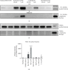
Figure 1
Figure 1
Effects of Nrf2 activators on Nrf2 expression in endothelial cells. A representative Western blot analysis of Nrf2 expression in nuclear (a, d) and cytoplasmic (b) fraction lysates obtained from HMEC-1 cells treated for 3 hours with 300 nM and 3 μM of bardoxolone methyl (Bm), dimethyl fumarate (Dmf), L-sulforaphane (L-s), culture medium alone, or with the addition of 0.05% DMSO. The expression of Lamin A/C was also determined to define fraction purity and serve as loading control (c). The results (d) are presented as a % of control (DMSO 0.05%) ± SEM, n = 3. The significance of the differences between the means was evaluated by one-way analysis of variance (ANOVA) with Dunnett's post hoc test; p < 0.05 and ∗∗∗∗p < 0.0001 indicate significant difference vs. the control group (DMSO 0.05%). The results were obtained in three independent experiments. In all panels, the first lane on the right contains a protein molecular weight marker.
Figure 2
Figure 2
Effects of Nrf2 activators on reactive oxygen species production in endothelial cells. HMEC-1 cells were incubated for 24 hours with bardoxolone methyl (Bm), dimethyl fumarate (Dmf), and L-sulforaphane (L-s), (300 nM and 5 μM). Intensity values of DHE fluorescence are expressed as relative fluorescence units ± SEM, n = 3. The significance of the differences between the means was evaluated by one-way analysis of variance (ANOVA) with Dunnett's post hoc test; ∗∗p < 0.01 and ∗∗∗∗p < 0.0001 indicate significant difference vs. the control group (DMSO 0.05%). The results were obtained in three independent experiments.
Figure 3
Figure 3
Effects of Nrf2 activators on endothelial viability assessed by the MTS test and cell counting. HMEC-1 cells were incubated with bardoxolone methyl, dimethyl fumarate, and L-sulforaphane (100 nM-50 μM) for 3 (a–c) and 24 (d–f) hours for the MTS test. Concentrations ranging from 100 nM up to 1 mM were applied in the cell counting test (g–i). Results are expressed as a % of control (cells treated with 0.05% DMSO). Values are expressed as mean ± SEM, n = 4. The significance of the differences between the means was evaluated by one-way analysis of variance (ANOVA) with Duncan's (MTS) or Dunnett's (cell counting) post hoc test if appropriate; p < 0.05, ∗∗p < 0.01, and ∗∗∗∗p < 0.0001 indicate significant difference vs. the control group (DMSO 0.05%). The results were obtained in four independent experiments.
Figure 4
Figure 4
Effects of Nrf2 activators on mitochondrial function in endothelial cells. HMEC-1 cells were incubated for three hours with bardoxolone methyl (a), dimethyl fumarate (b), and L-sulforaphane (c) (100 nM–5 μM). Values are expressed as mean oxygen consumption rate (OCR, in pmol/min ± SEM, n = 5). The significance of the differences between the means was evaluated by one-way analysis of variance (ANOVA) with Dunnett's post hoc test if appropriate; p < 0.05, ∗∗p < 0.01, and ∗∗∗p < 0.001 indicate significant difference vs. the control group (DMSO 0.05%). The results were obtained in five independent experiments.
Figure 5
Figure 5
Effects of Nrf2 activators on mitochondrial membrane potential in endothelial cells. HMEC-1 cells were incubated for three hours with FCCP (100 μM) or with bardoxolone methyl, dimethyl fumarate, and L-sulforaphane (100 nM–5 μM) (a). Values are expressed as a ratio of red and green fluorescence intensity of JC-1 calculated as a % of control ± SEM, n = 3. The significance of the differences between the means was evaluated by one-way analysis of variance (ANOVA) with Duncan's post hoc test if appropriate; p < 0.05 and ∗∗p < 0.01 indicate significant difference vs. the control group (DMSO 0.05%). (b) Representative images of HMEC-1 cells treated with 0.05% DMSO (upper panel), 5 μM of bardoxolone methyl (middle panel), and 100 μM of FCCP (lower panel). Images were collected with the use of 20x magnification at 561/617 nm (red fluorescence, JC-1 aggregates), 488/525 nm (green fluorescence, JC-1 monomers), and merged. The results were obtained in three independent experiments.
Figure 6
Figure 6
Effects of Nrf2 activators on secretion of endothelin-1 from endothelium to culture media. HMEC-1 cells were incubated for 3 (a) and 24 (b) hours with bardoxolone methyl, dimethyl fumarate, and L-sulforaphane (100 nM–5 μM). Values are expressed as mean ± SEM, n = 3. The significance of the differences between the means was evaluated by one-way analysis of variance (ANOVA) with Dunnett's post hoc test if appropriate; p < 0.0001 indicates significant difference vs. the control group (DMSO 0.05%). The results were obtained in three independent experiments.
Figure 7
Figure 7
Effects of endothelin-1 and endothelin-1 receptor antagonists on endothelial permeability. The measurement of impedance was performed using an ECIS assay at a frequency of 16,000 Hz in HMEC-1 cells: (a) incubated for one hour with medium, DMSO 0.05%, histamine 10 μM, and forskolin 5 μM; (b and c) incubated for three hours with endothelin-1 (ET-1) (100 pM–1000 pM); (d) incubated for three hours with 1 μM of BQ123 and/or BQ788. Values are expressed as mean normalized impedance ± SEM, n = 3. The significance of the differences between the means was evaluated by one-way analysis of variance (ANOVA) with Dunnett's post hoc test if appropriate; p < 0.05, ∗∗p < 0.01, ∗∗∗p < 0.001, and ∗∗∗∗p < 0.0001 indicate significant difference vs. the control groups: medium (a) and DMSO 0.05% (b, c, and d). The results were obtained in three independent experiments.
Figure 8
Figure 8
Effects of Nrf2 activators on endothelial permeability. The measurement of impedance was performed using an ECIS assay at a frequency of 16,000 Hz in HMEC-1 cells incubated for 3 and 24 hours with bardoxolone methyl (a and d), dimethyl fumarate (b and e), or L-sulforaphane (100 nM–5 μM) (c and f). Values are expressed as mean normalized impedance ± SEM, n = 3. The significance of the differences between the means was evaluated by one-way analysis of variance (ANOVA) with Duncan's post hoc test if appropriate; p < 0.05, ∗∗p < 0.01, and ∗∗∗∗p < 0.0001 indicate significant difference vs. the control group (DMSO 0.05%). The results were obtained in three independent experiments.

Similar articles

Cited by

References

    1. Suzuki T., Motohashi H., Yamamoto M. Toward clinical application of the Keap 1-Nrf2 pathway. Trends in Pharmacological Sciences. 2013;34(6):340–346. doi: 10.1016/j.tips.2013.04.005. - DOI - PubMed
    1. Malhotra D., Portales-Casamar E., Singh A., et al. Global mapping of binding sites for Nrf2 identifies novel targets in cell survival response through ChiP-Seq profiling and network analysis. Nucleic Acids Research. 2010;38(17):5718–5734. doi: 10.1093/nar/gkq212. - DOI - PMC - PubMed
    1. Baird L., Dinkova-Kostova A. T. The cytoprotective role of the Keap1–Nrf2 pathway. Archives of Toxicology. 2011;85(4):241–272. doi: 10.1007/s00204-011-0674-5. - DOI - PubMed
    1. Copple I. M. Current Concepts in Drug Metabolism and Toxicology. Elsevier; 2012. The Keap1–Nrf2 cell defense pathway—a promising therapeutic target? pp. 43–79. - DOI - PubMed
    1. Mandal A., Bishayee A. Trianthema portulacastrum Linn. displays anti-inflammatory responses during chemically induced rat mammary tumorigenesis through simultaneous and differential regulation of NF-κB and Nrf2 signaling pathways. International Journal of Molecular Sciences. 2015;16(2):2426–2445. doi: 10.3390/ijms16022426. - DOI - PMC - PubMed

MeSH terms

LinkOut - more resources