. 2020 Oct 17:2020:6427284.
doi: 10.1155/2020/6427284. eCollection 2020.

Ameliorating Effect of Klotho Protein on Rat Heart during I/R Injury

Affiliations

Ameliorating Effect of Klotho Protein on Rat Heart during I/R Injury

Agnieszka Olejnik et al. Oxid Med Cell Longev. .

Erratum in

Abstract

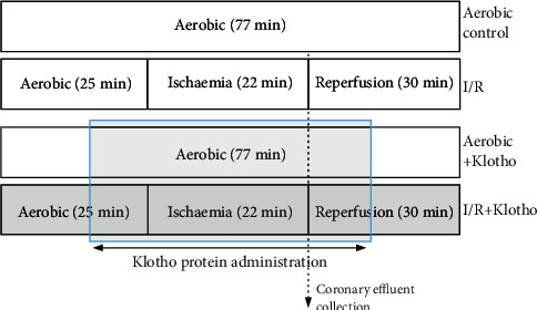
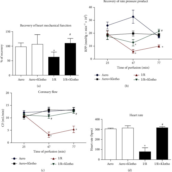
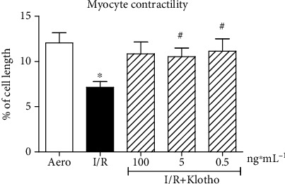
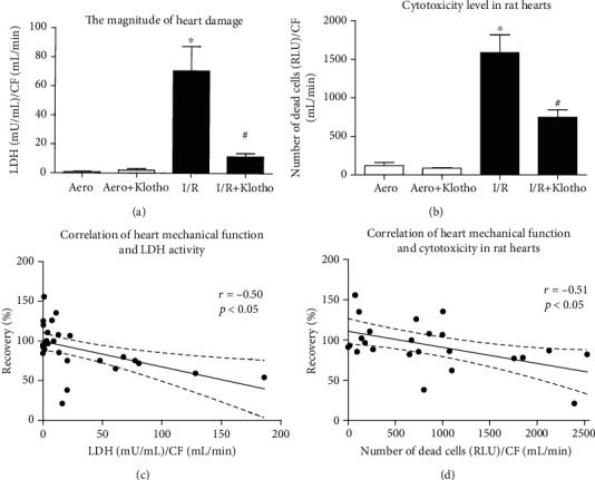
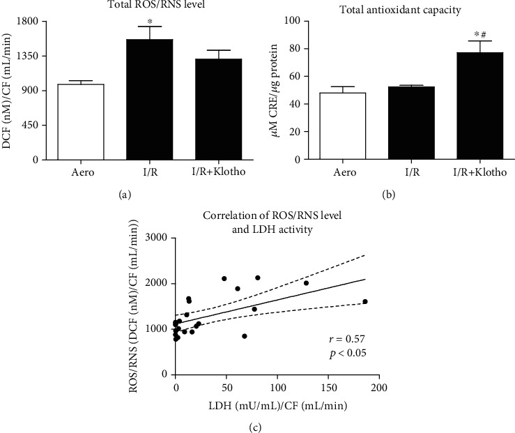
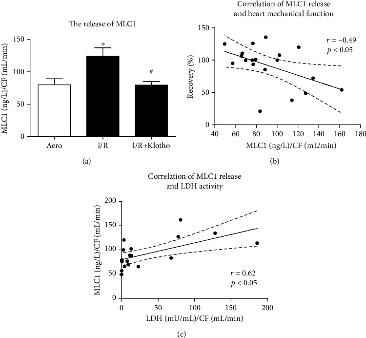
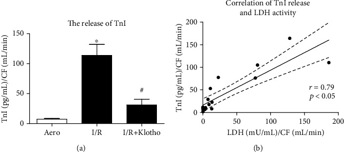
An essential procedure for the treatment of myocardial infarction is restoration of blood flow in the obstructed infarct artery, which may cause ischaemia/reperfusion (I/R) injury. Heart I/R injury manifests in oxidative stress, metabolic and morphological disorders, or cardiac contractile dysfunction. Klotho protein was found to be produced in the heart tissue and participate in antioxidation or ion homeostasis. The aim of this study was to examine an influence of Klotho protein on the heart subjected to I/R injury. Wistar rats served as a surrogate heart model ex vivo. Rat hearts perfused using the Langendorff method were subjected to global no-flow ischaemia, and isolated rat cardiomyocytes underwent chemical I/R in vitro, with or without recombinant Klotho protein administration. Haemodynamic parameters of heart function, cell contractility, markers of I/R injury and oxidative stress, and the level of contractile proteins such as myosin light chain 1 (MLC1) and troponin I (TnI) were measured. The treatment of hearts subjected to I/R injury with Klotho protein resulted in a recovery of heart mechanical function and ameliorated myocyte contractility. This improvement was associated with decreased tissue injury, enhanced antioxidant capacity, and reduced release of MLC1 and TnI. The present research showed the contribution of Klotho to cardioprevention during I/R. Thus, Klotho protein may support the protection from I/R injury and prevention of contractile dysfunction in the rat heart.


Conflict of interest statement

The authors declare that there is no conflict of interest regarding the publication of this paper.

Figures

Figure 1
Figure 1
Experimental protocol for I/R injury of isolated rat hearts, with and without Klotho administration. Klotho protein was added into the perfusion buffer and administered to isolated hearts during the last 10 min of aerobic stabilization (prior to the global ischaemia) and within the first 10 min of reperfusion after global ischaemia. I/R: ischaemia/reperfusion.
Figure 2
Figure 2
An effect of global I/R injury and Klotho protein on heart contractile function: (a) recovery of heart mechanical function. Percent recovery was calculated as a difference between RPP at 25 and 75 min of perfusion protocol; (b) RPP calculated as the product of the heart rate and pressure developed in the left ventricle (intraventricular pressure of left ventricle × heart rate/1000); (c) coronary flow; (d) heart rate at the end of reperfusion (77 min). Bpm: beats per minute; CF: coronary flow; I/R: ischaemia/reperfusion; RPP: rate pressure product; mean ± SEM; naero = 12; naero+Klotho = 3; nI/R = 14; nI/R+Klotho = 7; p < 0.05 vs. Aero; #p < 0.05 vs. I/R; ANOVA.
Figure 3
Figure 3
An influence of Klotho protein on isolated rat cardiomyocyte contractility. The contractility was expressed as peak shortening (%) in comparison to the length of the diastolic cell. Mean ± SEM; n = 6; p < 0.05 vs. Aero; #p < 0.05 vs. I/R; ANOVA.
Figure 4
Figure 4
An influence of Klotho on the magnitude of heart I/R injury: (a) the activity of LDH in coronary effluents as a marker of heart injury. LDH activity was normalized to CF; (b) the number of dead cells in rat hearts based on the activity of dead cell protease, tested by cytotoxicity assay. The data was expressed in RLU and normalized to CF; (c) correlation of heart mechanical function and LDH activity; (d) correlation of heart mechanical function and cytotoxicity in rat hearts. CF: coronary flow; LDH: lactate dehydrogenase; mU/mL: milli-international enzyme units per millilitre; RLU: relative light units; mean ± SEM; naero = 9; naero+Klotho = 3; nI/R = 10; nI/R+Klotho = 7; p < 0.05 vs. Aero; #p < 0.05 vs. I/R; ANOVA.
Figure 5
Figure 5
Oxidative status in the hearts subjected to I/R injury: (a) total ROS and RNS level expressed as nM of DCF and normalized to coronary flow; (b) total antioxidant capacity of hearts subjected to I/R. TAC was expressed as μM of CRE and normalized to total protein concentration; (c) correlation of ROS/RNS level and LDH activity. CF: coronary flow; CRE: copper reducing equivalents; DCF: 2′, 7′-dichlorodihydrofluorescein; LDH: lactate dehydrogenase; mU/mL: milli international enzyme units per millilitre; RNS: reactive nitrogen species; ROS: reactive oxygen species; TAC: total antioxidant capacity; mean ± SEM; naero = 8; nI/R = 9; nI/R+Klotho = 6; p < 0.05 vs. Aero; #p < 0.05 vs. I/R; ANOVA.
Figure 6
Figure 6
An influence of Klotho protein on MLC1 amount in rat hearts: (a) concentration of MLC1 in coronary effluents tested by ELISA and normalized to CF; (b) correlation of MLC1 concentration and heart mechanical function; (c) correlation of MLC1 concentration and LDH activity. CF: coronary flow; GAPDH: glyceraldehyde 3-phosphate dehydrogenase; LDH: lactate dehydrogenase; mU/mL: milli-international enzyme units per millilitre; MLC1: myosin light chain 1; mean ± SEM; naero = 7; nI/R = 5; nI/R+Klotho = 7; p < 0.05 vs. Aero; #p < 0.05 vs. I/R; ANOVA.
Figure 7
Figure 7
An influence of Klotho protein on TnI release in rat hearts: (a) concentration of TnI in coronary effluents tested by ELISA and normalized to CF; (b) correlation of TnI concentration and LDH activity. CF: coronary flow; LDH: lactate dehydrogenase; mU/mL: milli-international enzyme units per millilitre; TnI: troponin I; mean ± SEM; naero = 7; nI/R = 5; nI/R+Klotho = 7; p < 0.05 vs. Aero; #p < 0.05 vs. I/R; ANOVA.

Similar articles

Cited by

References

    1. Antman E. M., Anbe D. T., Armstrong P. W., et al. ACC/AHA guidelines for the management of matients with ST-elevation myocardial infarction—executive summary: a report of the American College of Cardiology/American Heart Association Task Force on Practice Guidelines (writing committee to revise the 1999 guidelines for the management of patients with acute myocardial infarction) Circulation. 2004;110:588–636. - PubMed
    1. Heusch G., Gersh B. J. The pathophysiology of acute myocardial infarction and strategies of protection beyond reperfusion: a continual challenge. European Heart Journal. 2017;38(11):774–784. doi: 10.1093/eurheartj/ehw224. - DOI - PubMed
    1. Tsutsui H., Kinugawa S., Matsushima S. Oxidative stress and heart failure. American Journal of Physiology. Heart and Circulatory Physiology. 2011;301(6):H2181–H2190. doi: 10.1152/ajpheart.00554.2011. - DOI - PubMed
    1. Kuro-o M. Klotho as a regulator of fibroblast growth factor signaling and phosphate/calcium metabolism. Current Opinion in Nephrology and Hypertension. 2006;15(4):437–441. doi: 10.1097/01.mnh.0000232885.81142.83. - DOI - PubMed
    1. Kuro-o M., Matsumura Y., Aizawa H., et al. Mutation of the mouse klotho gene leads to a syndrome resembling ageing. Nature. 1997;390(6655):45–51. doi: 10.1038/36285. - DOI - PubMed

MeSH terms

LinkOut - more resources