. 2020 Oct 5:10:581814.
doi: 10.3389/fonc.2020.581814. eCollection 2020.

Mifepristone as a Potential Therapy to Reduce Angiogenesis and P-Glycoprotein Associated With Glioblastoma Resistance to Temozolomide

Affiliations

Mifepristone as a Potential Therapy to Reduce Angiogenesis and P-Glycoprotein Associated With Glioblastoma Resistance to Temozolomide

Monserrat Llaguno-Munive et al. Front Oncol. .

Erratum in

Abstract

Glioblastoma, the most common primary central nervous system tumor, is characterized by extensive vascular neoformation and an area of necrosis generated by rapid proliferation. The standard treatment for this type of tumor is surgery followed by chemotherapy based on temozolomide and radiotherapy, resulting in poor patient survival. Glioblastoma is known for strong resistance to treatment, frequent recurrence and rapid progression. The aim of this study was to evaluate whether mifepristone, an antihormonal agent, can enhance the effect of temozolomide on C6 glioma cells orthotopically implanted in Wistar rats. The levels of the vascular endothelial growth factor (VEGF), and P-glycoprotein (P-gp) were examined, the former a promoter of angiogenesis that facilitates proliferation, and the latter an efflux pump transporter linked to drug resistance. After a 3-week treatment, the mifepristone/temozolomide regimen had decreased the level of VEGF and P-gp and significantly reduced tumor proliferation (detected by PET/CT images based on 18F-fluorothymidine uptake). Additionally, mifepristone proved to increase the intracerebral concentration of temozolomide. The lower level of O6-methylguanine-DNA-methyltransferase (MGMT) (related to DNA repair in tumors) previously reported for this combined treatment was herein confirmed. After the mifepristone/temozolomide treatment ended, however, the values of VEGF, P-gp, and MGMT increased and reached control levels by 14 weeks post-treatment. There was also tumor recurrence, as occurred when administering temozolomide alone. On the other hand, temozolomide led to 100% mortality within 26 days after beginning the drug treatment, while mifepristone/temozolomide enabled 70% survival 60-70 days and 30% survived over 100 days, suggesting that mifepristone could possibly act as a chemo-sensitizing agent for temozolomide.

Keywords: P-gp; angiogenesis; drug resistance; glioblastoma; mifepristone; temozolomide.


Figures

FIGURE 1
FIGURE 1
Tumor growth in the orthotopic rat model of glioma was evaluated by comparing animals weight between groups: negative control (formula image
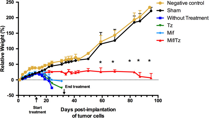
) and sham surgery (formula image); and in four groups with implanted glioma cancer cells, one without drug treatment (formula image) and the other given temozolomide only (Tz) (formula image), mifepristone only (Mif) (formula image), and mifepristone/temozolomide (Mif/Tz) (formula image). Each point of the graphic represents the mean ± SEM of six animals. *Significant difference (p < 0.05) between Mif/Tz and sham.
FIGURE 2
FIGURE 2
Hematoxylin and eosin (H&E) staining analysis of glioma tissue. Hyperbasophilic cells (black arrow), hyperchromatics cells (red arrow), vessel proliferation (arrowhead), mitosis (blue arrow). The images are representative of three animals per treatment Scale bars = 50 μm.
FIGURE 3
FIGURE 3
(A) Immunohistochemical staining of CD31 marker. Vessel density was assessed by immunostaining for CD31 positive glioma cell nuclei in rats implanted with glioma. The images are representative of three animals per treatment. Scale bar 50 μm. (B) Expression of VEGF at the end of the 3-week drug treatment, showing a significantly lower level in the mifepristone/temozolomide (Mif/Tz)-treated group versus the untreated (W/T) group, both with implanted cancer cells. Data are expressed as the mean ± SD from eight independent experiments. *Significant difference (p < 0.05) between the Mif/Tz and W/T group.
FIGURE 4
FIGURE 4
The quantification of P-gp levels at the end of the 3-week drug treatment evidenced a significant downregulation in the rats given mifepristone (Mif) or mifepristone/temozolomide (Mif/Tz) compared to those receiving no drug treatment (W/T) or temozolomide (Tz). (A) Representative Western blot; h.e., high exposure; l.e., low exposure. (B) densitometric analysis of the P-gp protein. Data are expressed as the mean ± SD from three independent experiments. *Significant difference (p < 0.05).
FIGURE 5
FIGURE 5
(A) Based on typical chromatograms of temozolomide in brain tissue, there was a significantly higher concentration of temozolomide (Tz) in rats given a pre-treatment of 60 mg/kg of mifepristone (Mif) followed by 30 mg/kg of Tz (red line) than in animals receiving only 30 mg/kg of Tz (green line). (B) Bar graph illustration of the Tz uptake in rat brain tissue (n = 6 ± SD). *Significant difference (p < 0.05).
FIGURE 6
FIGURE 6
Proliferative activity in the orthotopic model of glioma evaluated by PET/CT images showing tumor uptake of 18F-FLT. (A) The images reveal the relative tumor size at 5, 7, 9, and 14 weeks post-surgery. Drug treatments were given from weeks 2–5. (B) The activity proliferative of tumors measured as total proliferation (TLP). (C) Survival analysis for 100 days after tumor cells implantation.
FIGURE 7
FIGURE 7
Hematoxylin and eosin (H&E) staining analysis of glioma tissue. The images are representative of three animals per treatment. Hyperbasophilic cells (black arrow), Hyperchromatics cells (red arrow), vessel proliferation (arrowhead), mitosis (blue arrow). Scale bars = 50 μm.
FIGURE 8
FIGURE 8
Effect of mifepristone/temozolomide on VEGF during tumor recurrence. (A) Immunohistochemical stainings with CD31 marker. Vessel density was assessed by immunostaining for CD31 positive glioma cell nuclei in rats implanted with glioma. The images are representative of three animals per treatment. Scale bar 50 μm. (B) Expression of VEGF in the sham-operated rats, implant-operated animals with no drug treatment (W/T), and at 5, 9 and 14 weeks post-surgery (the mifepristone/temozolomide (Mif/Tz) treatment were given only during weeks 2–5). Compared to the W/T rats, the Mif/Tz animals showed a lower level of VEGF at 5 weeks and a similar level at the time of tumour recurrence, at 9 and 14 weeks post-surgery. Data are expressed as the mean ± SD from five independent experiments. * Significant difference (p < 0.05) between the Mif/Tz and W/T group.
FIGURE 9
FIGURE 9
(A) Comparison of the levels of P-gp (determined by Western Blot) in the sham-operated rats and two groups of implant-operated animals: one with no drug treatment (W/T) and the other given mifepristone/temozolomide (Mif/Tz) at 5 weeks post-surgery (corresponding to the end of the 3-week drug treatment), and 9, 14 weeks (corresponding to 4 and 9 weeks after the end of drug treatment), h.e., high exposure; l.e., low exposure. (B) The densitometer analysis (n = 3). Data represent the mean ± SD of three independent experiments. *Significant difference (p < 0.05) between the Mif/Tz rats at 5 and 14 weeks.
FIGURE 10
FIGURE 10
(A) Comparison of the level of the DNA repair enzyme, MGMT, determined by Western blot in the sham-operated rats and two groups of implant-operated animals: one with no drug treatment (W/T) and the other given mifepristone/temozolomide (Mif/Tz) at 5 weeks post-surgery (corresponding to the end of the 3-week drug treatment) and 9 and 14 weeks after surgery. A lower level of MGMT was found in the Mif/Tz versus W/T group at 5 weeks post-surgery, an effect that was gradually reversed. (B) Densitometer analysis (n = 3). Data is expressed as the mean ± SD of three independent experiment. * Significant difference (p < 0.05).
FIGURE 11
FIGURE 11
Schematic portrayal of the possible mechanisms of the combination mifepristone/temozolomide treatment that improved the effect found with temozolomide alone. The mechanisms studied were: (1) the inhibition of angiogenesis, measured as reduced levels of VEGF; (2) the attenuation of DNA repair, evaluated as a decrease in MGMT; and (3) the increased capacity of temozolomide to pass through the BBB, assessed as a lower P-gp level and a higher concentration of temozolomide in brain cells. As described in a previous report by our group (5), mifepristone diminishes the level of anti-apoptotic protein Bcl-2 and impedes endothelial cell survival in tumors. This may be the mechanism by which mifepristone/temozolomide herein lowered the level of VEGF. The treatment with mifepristone or temozolomide alone decreased the levels of VEGF to a lesser extent, perhaps by the blockade of autocrine VEGF signaling through specific down-regulation of NRP-1. Additionally, a decline in the expression P-gp was found when administering mifepristone/temozolomide. Thus, this combination treatment may allow for an enhanced intratumoral concentration of temozolomide and contribute to greater tumor cell death. The latter was evidenced by lower tumor proliferation during the drug treatment period. As can be appreciated, mifepristone appears to sensitize glioblastoma cells to the effects of temozolomide.

Similar articles

Cited by

References

    1. Johnson DR, O’Neill BP. Glioblastoma survival in the United States before and during the temozolomide era. J Neurooncol. (2012) 107:359–64. 10.1007/s11060-011-0749-4 - DOI - PubMed
    1. Thakkar JP, Dolecek TA, Horbinski C, Ostrom QT, Lightner DD, Barnholtz-Sloan JS, et al. Epidemiologic and molecular prognostic review of glioblastoma. Cancer Epidemiol Biomarkers Prev. (2014) 23:1985–96. 10.1158/1055-9965.EPI-14-0275 - DOI - PMC - PubMed
    1. Witthayanuwat S, Pesee M, Supaadirek C, Supakalin N, Thamronganantasakul K, Krusun S. Survival analysis of glioblastoma multiforme. Asian Pac J Cancer Prev. (2018) 19:2613–7. 10.22034/APJCP.2018.19.9.2613 - DOI - PMC - PubMed
    1. Wang H, Xu T, Jiang Y, Xu H, Yan Y, Fu D, et al. The challenges and the promise of molecular targeted therapy in malignant gliomas. Neoplasia. (2015) 17:239–55. 10.1016/j.neo.2015.02.002 - DOI - PMC - PubMed
    1. Llaguno-Munive M, Romero-Pina M, Serrano-Bello J, Medina LA, Uribe-Uribe N, Salazar AM, et al. Mifepristone overcomes tumor resistance to temozolomide associated with DNA damage repair and apoptosis in an orthotopic model of glioblastoma. Cancers. (2019) 11:16. 10.3390/cancers11010016 - DOI - PMC - PubMed