. 2020 Oct 15:2020:7930160.
doi: 10.1155/2020/7930160. eCollection 2020.

Double-Targeted Knockdown of miR-21 and CXCR4 Inhibits Malignant Glioma Progression by Suppression of the PI3K/AKT and Raf/MEK/ERK Pathways

Affiliations

Double-Targeted Knockdown of miR-21 and CXCR4 Inhibits Malignant Glioma Progression by Suppression of the PI3K/AKT and Raf/MEK/ERK Pathways

Feijiao Liu et al. Biomed Res Int. .

Abstract

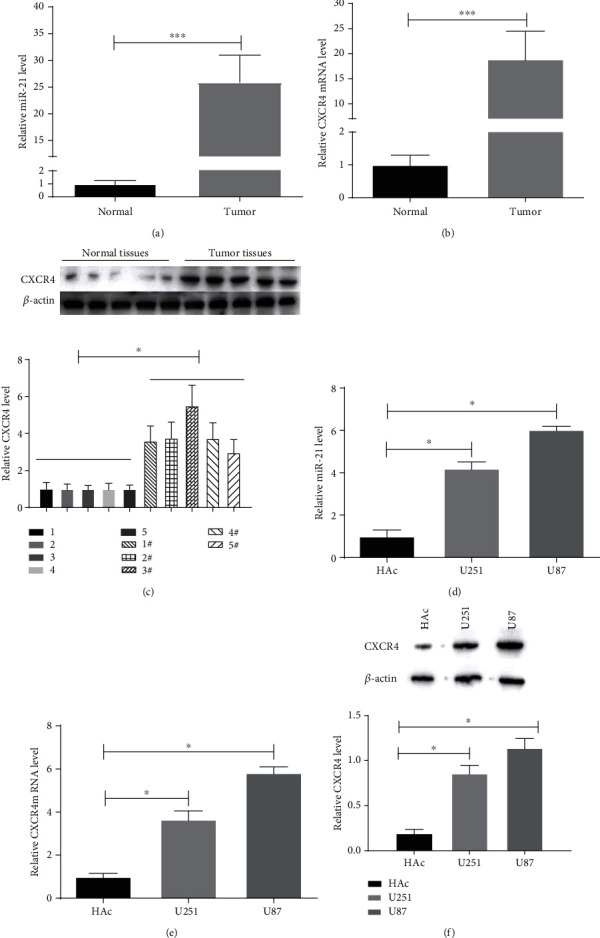
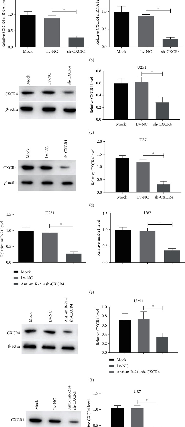
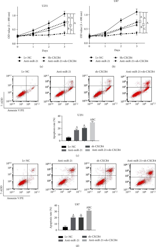
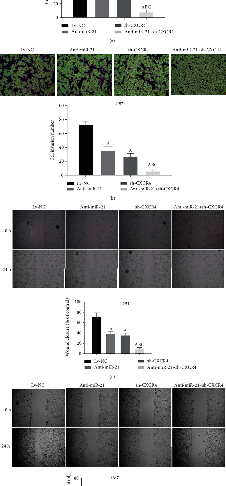
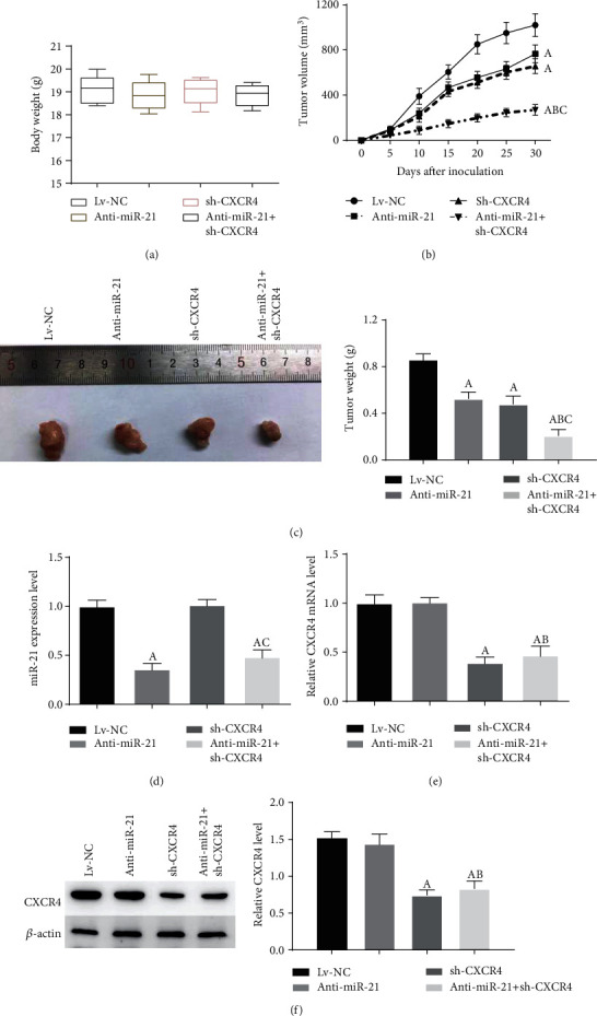
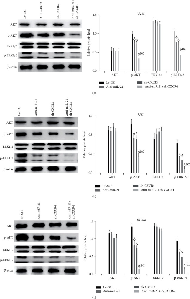
Currently, miR-21 and CXCR4 are being extensively investigated as two key regulators in glioma malignancy. In this study, we investigated the combined effects of these two factors on glioma progression. Herein, the expression of miR-21 and CXCR4 was increased in tumor tissues and cell lines. Inhibition of miR-21, CXCR4, and miR-21 and CXCR4 together all reduced the migration, invasiveness, proliferation, and enhanced apoptosis in glioma cells, as well as reduced tumor volume and mass in xenograft model. The inhibition effect was strongest in double-targeted knockdown of miR-21 and CXCR4 group, whose downstream pathways involved in AKT axis and ERK axis activation. In conclusion, our findings reported that double-targeted knockdown of miR-21 and CXCR4 could more effectively inhibit the proliferation, migration, invasion, and growth of transplanted tumor and promote cell apoptosis, which were involved in the PI3K/AKT and Raf/MEK/ERK signaling pathways.


Conflict of interest statement

No potential conflicts of interest were disclosed.

Figures

Figure 1
Figure 1
miR-21 and CXCR4 levels in malignant glioma tissues and cells. (a, b) The expression of miR-21 and CXCR4 was detected by qRT-PCR in glioma and normal adjacent tissues samples. (c) The levels of CXCR4 were determined by western blot in tumor and normal tissues samples. (d, e) The expression of miR-21 and CXCR4 was measured by qRT-PCR in glioma cells (U251 and U87) and control cells (HAc). (f) CXCR4 protein levels were tested by western blot. P < 0.05.
Figure 2
Figure 2
miR-21 and CXCR4 levels of stable glioma cells transfected with anti-miR-21, sh-CXCR4, or anti-miR-21 + sh-CXCR4. (a, b) miR-21 and CXCR4 levels in the transfected U251 and U87 cells were measured using qRT-PCR. (c, d) CXCR4 protein levels in the transfected U251 and U87 cells were measured using western blot. (e) miR-21 and CXCR4 levels in the transfected U251 and U87 cells were measured using qRT-PCR. (f, g) CXCR4 protein levels in the transfected U251 and U87 cells were measured using western blot. P < 0.05.
Figure 3
Figure 3
Double-targeted knockdown of miR-21 and CXCR4 inhibited proliferation of glioma cells. (a, b) MTT assays were employed to assess the proliferation of transfected U251 and U87 cells. (c, d) Flow cytometry was used to analyze the apoptosis in transfected U251 and U87 cells. P < 0.05, (A) vs. Lv-NC group; (B) vs. anti-miR-21 group; (C) vs. sh-CXCR4 group.
Figure 4
Figure 4
Double-targeted knockdown of miR-21 and CXCR4 inhibited invasion and migration of glioma cells. (a, b) Transwell assay was performed on the transfected U251 and U87 cells. Magnification, 200x. (c, d) Scratch assays were performed to detect migration of transfected cells. Magnification, 40x. P < 0.05, (A) vs. Lv-NC group; (B) vs. anti-miR-21 group; (C) vs. sh-CXCR4 group.
Figure 5
Figure 5
Double-targeted knockdown of miR-21 and CXCR4 inhibited glioma xenograft growth. (a) Mice were weighted at 30 day postinoculation. (b) Tumor volumes were monitored for 30 days. (c) Tumors were dissected and weighted. (d, e) miR-21 and CXCR4 levels in xenograft tumor tissues were measured using qRT-PCR. (f) CXCR4 protein levels were tested using western blot. P < 0.05, (A) vs. Lv-NC group; (B) vs. anti-miR-21 group; (C) vs. sh-CXCR4 group.
Figure 6
Figure 6
Double-targeted knockdown of miR-21 and CXCR4 inhibited the PI3K/AKT and Raf/MEK/ERK pathways in glioma xenograft tissues and cells. (a–c) Related proteins were tested using western blot. P < 0.05, (A) vs. Lv-NC group; (B) vs. anti-miR-21 group; (C) vs. sh-CXCR4 group.

Similar articles

Cited by

References

    1. Yang X. J., Long H. A., Sheng-ping Y. U. Study on invasion and migration of malignant glioma. Chinese Journal of Contemporary Neurology & Neurosurgery. 2018;18(1):p. 36.
    1. Sottoriva A., Spiteri I., Piccirillo S. G. M., et al. Intratumor heterogeneity in human glioblastoma reflects cancer evolutionary dynamics. Proceedings of the National Academy of Sciences of United States of America. 2013;110(10):4009–4014. doi: 10.1073/pnas.1219747110. - DOI - PMC - PubMed
    1. Skalsky R. L., Cullen B. R. Reduced expression of brain-enriched microRNAs in glioblastomas permits targeted regulation of a cell death gene. PLoS One. 2011;6(9, article e24248) doi: 10.1371/journal.pone.0024248. - DOI - PMC - PubMed
    1. Põlajeva J., Swartling F. J., Jiang Y., et al. miRNA-21 is developmentally regulated in mouse brain and is co-expressed with SOX2 in glioma. BMC Cancer. 2012;12(1) doi: 10.1186/1471-2407-12-378. - DOI - PMC - PubMed
    1. Lei B. X., Liu Z. H., Li Z. J., Li C., Deng Y. F. miR-21 induces cell proliferation and suppresses the chemosensitivity in glioblastoma cells via downregulation of FOXO1. International Journal of Clinical and Experimental Medicine. 2014;7(8):2060–2066. - PMC - PubMed

MeSH terms