. 2021 May;70(5):1189-1202.
doi: 10.1007/s00262-020-02764-9. Epub 2020 Oct 29.

Impact of tumor heterogeneity and microenvironment in identifying neoantigens in a patient with ovarian cancer

Affiliations

Impact of tumor heterogeneity and microenvironment in identifying neoantigens in a patient with ovarian cancer

Tao Dao et al. Cancer Immunol Immunother. 2021 May.

Abstract

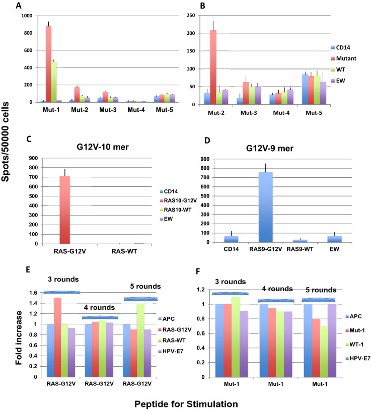
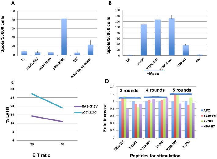
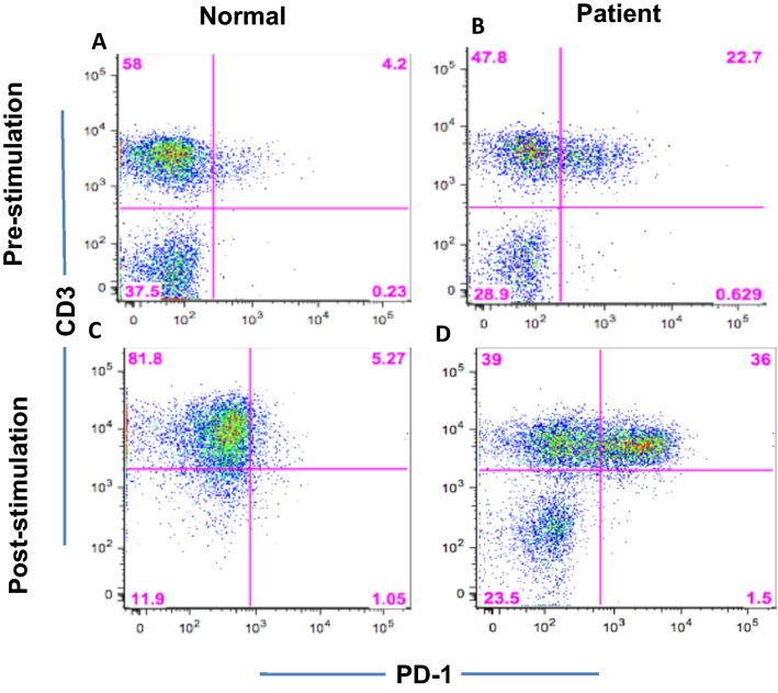
Identification of neoepitopes as tumor-specific targets remains challenging, especially for cancers with low mutational burden, such as ovarian cancer. To identify mutated human leukocyte antigen (HLA) ligands as potential targets for immunotherapy in ovarian cancer, we combined mass spectrometry analysis of the major histocompatibility complex (MHC) class I peptidomes of ovarian cancer cells with parallel sequencing of whole exome and RNA in a patient with high-grade serous ovarian cancer. Four of six predicted mutated epitopes capable of binding to HLA-A*02:01 induced peptide-specific T cell responses in blood from healthy donors. In contrast, all six peptides failed to induce autologous peptide-specific response by T cells in peripheral blood or tumor-infiltrating lymphocytes from ascites of the patient. Surprisingly, T cell responses against a low-affinity p53-mutant Y220C epitope were consistently detected in the patient with either unprimed or in vitro peptide-stimulated T cells even though the patient's primary tumor did not bear this mutation. Our results demonstrated that tumor heterogeneity and distinct immune microenvironments within a patient should be taken into consideration for identification of immunogenic neoantigens. T cell responses to a driver gene-derived p53 Y220C mutation in ovarian cancer warrant further study.

Keywords: Immunotherapy; Mass spectrometry; Neoepitopes; Ovarian cancer; Parallel sequencing; T cell response.


Conflict of interest statement

David Scheinberg is on the advisory board of, has received consulting fees from, and/or has equity in Progenics Pharmaceuticals, Sellas, KLUS, Iovance Biotherapeutics, Inc., Pfizer, Actinium Pharmaceuticals, Inc., OncoPep, and Eureka Therapeutics. Tao Dao has equity in Eureka Therapeutics. Dr O'Cearbhaill's institution receives funding for clinical research that she is leading from Celgene/Juno, Tesaro/GSK, Ludwig Cancer Institute, Abbvie, Regeneron, TCR2 Therapeutics, Atara Biotherapeutics, MarkerTherapeutics, Syndax Pharmaceuticals, Genmab Therapeutics, Sellas Therapeutics, Genentech, Kite Pharma, and the Gynecologic Oncology Foundation. Dr O’Cearbhaill has served on a once-off advisory board for Regeneron, Genmab Therapeutics, and GSK. All other authors declare no conflict or competing interests.

Figures

Fig. 1
Fig. 1
Mutant peptide-induced T cell response in HLA-A*02:01+ healthy donors. a CD3 T cells from HLA-A0*02:01 donor were stimulated with five newly identified HLA-A2-binding peptides (listed in Table 1) for three rounds, and T cell response was measured against the mutant peptides, their wild-type (WT) peptides, or with irrelevant HLA-A2-binding peptide EW presented by autologous CD14+ cells by IFN-γ ELISPOT assay. CD14+ antigen-presenting cells (APCs) alone served as a negative control. b CD3 T cells from an HLA-A0*02:01+ donor were stimulated for up to four rounds with four peptides and tested for IFN-γ secretion in the same manner as described in Panel A. c T cells from an HLA-A*02:01+ donor were stimulated for three rounds with RAS-G12V-mutant peptide (10mer) or its native sequence (RAS-WT), and the peptide-specific T cell response was measured by IFN-γ ELISPOT assay. d Similarly, T cells from an HLA-A*02:01+ donor were stimulated for three rounds with RAS-G12V-mutant peptide (9mer), and the peptide-specific T cell response was measured by IFN-γ ELISPOT assay. For panels A to D, each point represents average ± SD from triplicate cultures. Data represent results from five to six similar experiments from multiple donors. T cells of peripheral blood mononuclear cells (PBMCs) from the patient were stimulated with RAS-G12V or mutant #1 peptide for three, four, and five rounds, and peptide-specific responses were measured by IFN-γ ELISPOT assay. Spot numbers were normalized by fold increase over the T cells cultured with only APCs, without peptides (e and f). The data represent the average of triplicate micro-well cultures
Fig. 2
Fig. 2
p53 Y220C-mutant peptide-specific T cell response in a patient with high-grade serous ovarian cancer. a Spontaneous T cell response against p53 Y220C-mutant peptide. Ascites cells from the patient with ovarian cancer were cultured in 5% FBS-RPMI1640 medium for 7 days in the presence of IL-2 (10 unit) and IL-15 (5 ng/mL). The cells were harvested, washed, and tested against T2 cells pulsed with three HLA-A2-binding p53 mutant peptides, as indicated, and tumor cells were isolated from the ascites. T2 cells alone or pulsed with EW peptide were used as negative controls. b Ascites cells were stimulated with Y220C peptide for three rounds, and peptide-specific T cell response was measured against Y220C mutant with or without the presence of anti–programmed cell death protein 1 (PD-1) monoclonal antibody (mAb) or isotype control mAb (10 ug/mL) during the ELISPOT assay (overnight culture). Autologous dendritic cells (DCs), wild-type (Y220-WT) peptide, and EW served as controls. Each point represents average ± SD from triplicate cultures. c In parallel, cytotoxicity of T cells stimulated with RAS-G12V or Y220C peptides were tested against autologous tumor cells from the patient, measured by standard 51Cr release assay. Each point represents average ± SD from triplicate cultures. d T cells of PBMCs from the patient were stimulated with p53 Y220C or its native sequence (WT) for three, four, and five rounds, and the peptide-specific responses were measured by IFN-γ ELISPOT assay. Spot numbers were normalized by fold increase over the T cells cultured with only APCs, without peptides. The data represent the average of triplicate micro-well cultures
Fig. 3
Fig. 3
Programmed cell death protein 1 (PD-1) expression. Peripheral blood mononuclear cells (PBMCs) from a healthy donor (a) or lymphocytes isolated from the patient’s ascites (b) were tested for the PD-1 expression on CD3+ T cells prior to peptide stimulation. c, d The cells were stimulated with mutant #1 peptide for three rounds, and PD-1 expression was shown on CD3+ T cells (lower two panels). No PD-1 expression was found in CD19+ B cells and CD14+ macrophages (data not shown)
Fig. 4
Fig. 4
Programmed death-ligand 1 (PD-L1) expression. Dendritic cells (DCs) were generated from the patient and a healthy donor, as described in the Materials and Methods. PD-L1 expression was determined by staining the DCs with CD11C and PD-L1. CD11C+ cells were gated, and PD-L1 expression was compared between the patient and healthy donor by dot plot and histogram overlay (a and b) (Green and orange: isotype control and PD-L1 expression on CD11C+ DCs from the patient. Red and blue: isotype control and PD-L1 expression on CD11C+ DCs from a healthy donor). Tumor cells isolated from the patient’s ascites were tested for the expression of tumor markers HER-2, EpCAM and MUC16 (ce). PD-L1 expression on MUC16-positive tumor cells was shown by overlay of dot plot staining with isotype control. Percentages in each quadrant and blue dots are MUC16 vs. PD-L1. The red dots in the lower left quadrant show isotype controls (negative) for both MUC16 and PD-L1 monoclonal antibodies (f)
Fig. 5
Fig. 5
IFN-γ secretion by T cells. Peripheral blood mononuclear cells (PBMCs) from healthy donor or lymphocytes isolated from patient were stimulated with RAS-G12V-mutant peptide (10mer) in the presence of monoclonal antibodies (mAbs) to human programmed cell death protein 1 (PD-1) (a) or isotype control (b) during the weekly priming of three rounds. Intracellular IFN-γ secretion by CD8+ T cells of the patient prior to (c) and after three rounds of stimulation with RAS-G12V-mutant peptide in the presence of anti–PD-1 mAb (d) was shown. CD3 T cells from a healthy HLA-A*02:01+ donor prior to (e) and after three rounds of stimulation with RAS-G12V (f) in the presence of anti–PD-1 mAb was tested for Intracellular IFN-γ secretion, in parallel. The data represent one of three sets of stimulation

References

    1. Schumacher T, Schreiber RD. Neoantigens in cancer immunotherapy. Science. 2015;348:69–74. doi: 10.1126/science.aaa4971. - DOI - PubMed
    1. Luksza M, Riaz N, Makarov V, Balanchandran V, Hellmann MD, Solovyov A, et al. A neoantigens fitness model predicts tumor response to checkpoint blockade immunotherapy. Nature. 2017;551:517–520. doi: 10.1038/nature24473. - DOI - PMC - PubMed
    1. Tran E, Robbins PF, Rosenberg SA. “Final common pathway” of human cancer immunotherapy: targeting random somatic mutations. Nat Immunol. 2017;18:255–262. doi: 10.1038/ni.3682. - DOI - PMC - PubMed
    1. Tran E, Ahmadzadeh M, Lu YC, Gros A, Turcitte S, et al. Immunogenicity of somatic mutations in human gastrointestinal cancers. Science. 2015;350:1387–1390. doi: 10.1126/science.aad1253. - DOI - PMC - PubMed
    1. Carreno BM, Magrini V, Becker-Hapak M, Kaabinejadian S, Hundal J, Petti AA, et al. Cancer immunotherapy. A dendritic cell vaccine increases the breadth and diversity of melanoma noeantigen-specific T cells. Science. 2015;348:803–808. doi: 10.1126/science.aaa3828. - DOI - PMC - PubMed

MeSH terms