. 2021 Jun;99(4):e470-e479.
doi: 10.1111/aos.14642. Epub 2020 Oct 30.

Comprehensive analysis of genetic and clinical characteristics of 30 patients with X-linked juvenile retinoschisis in China

Affiliations

Comprehensive analysis of genetic and clinical characteristics of 30 patients with X-linked juvenile retinoschisis in China

Feng-Juan Gao et al. Acta Ophthalmol. 2021 Jun.

Abstract

Purpose: To provides the clinical and genetic characteristics of a series of Chinese patients with X-linked juvenile retinoschisis (XLRS) through multimodal imaging and next-generation sequencing.

Methods: Thirty patients (60 eyes) from 29 unrelated families of Chinese origin with XLRS were screened using multigene panel testing, and underwent a complete clinical evaluation. All variants identified in this study and reported in the Human Gene Mutation Database were analysed.

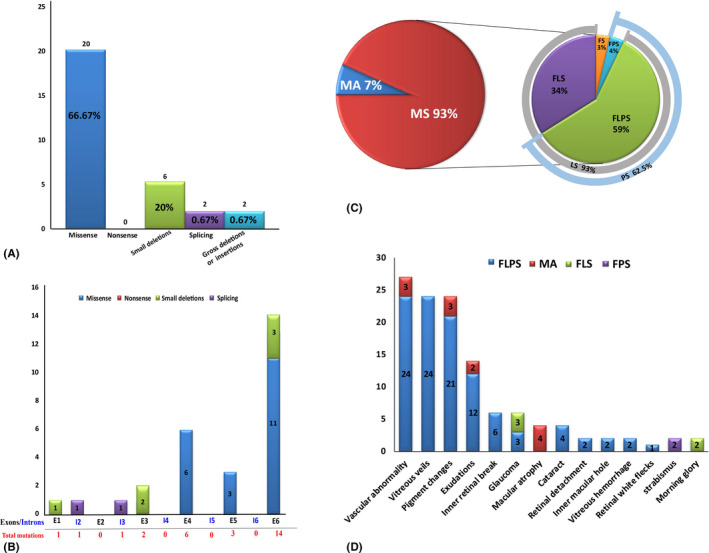
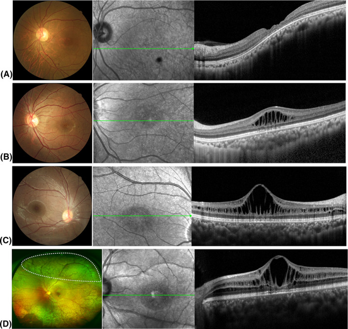
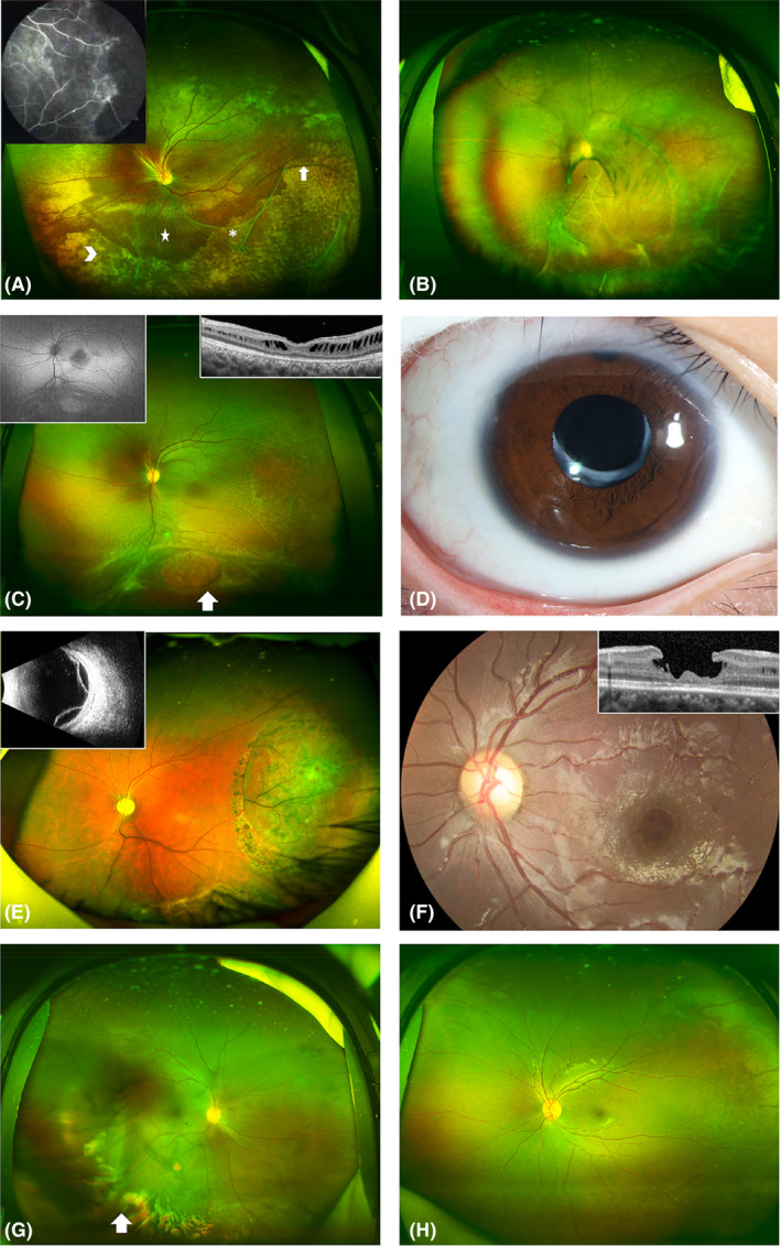
Results: Twenty-five distinct variants in the retinoschisin gene were identified, of which eight were novel, and one was de novo. Missense mutations were the most prevalent type, and mutation hot spot was localized in the discoidin domain. The mean Snellen best-corrected visual acuity was 0.28 ± 0.17. Of all eyes presenting with schisis, 92.86% had lamellar schisis and 62.5% had peripheral schisis. Schisis changes mostly involved inner and outer nuclear layers. X-linked juvenile retinoschisis (XLRS) patients had a high incidence of complications, and peripheral schisis was a risk factor for it. No obvious genotype-phenotype association was observed.

Conclusion: This study provides comprehensive analyses of the genetic and clinical characteristics of XLRS in a cohort of Chinese patients. The fourth de novo mutation in RS1 was identified. And we show that XLRS has a wide spectrum of clinical characteristics; hence, molecular diagnosis is crucial for its diagnosis, differential diagnosis and genetic counselling. Peripheral schisis is a risk factor for the high incidence of complications, and no clear genotype-phenotype correlations were found.

Keywords: RS1; Chinese population; X-linked retinoschisis; clinical diagnosis; molecular genetics; optical coherence tomography.


Figures

Fig. 1
Fig. 1
Overview of pathologic RS1 mutations identified in this study and complications of all the patients. (A) Number of RS1 mutations of different mutation types identified in this study. (B) Distribution of the RS1 variants identified in this study. Exons are numbered in black font, and introns are numbered in blue font. Total mutations are shown in red font. (C) Pie chart showing the structural changes in XLRS patients. Macular schisis was seen in 93% of patients, with lamellar schisis (LS) accounting for 93% and peripheral schisis (PS) accounting for 62.5%. (D) Distribution of the different complications observed in different types of patients. FLPS = complex type patients who had foveal, lamellar and peripheral schisis., FLS = foveo‐lamellar patients who had foveal schisis and lamellar schisis, but not peripheral schisis, FPS = foveo‐peripheral type patients who had foveal and peripheral schisis without lamellar schisis, FS = foveal schisis, MA = macular atrophy.
Fig. 2
Fig. 2
Representative fundus photographs and optical coherence tomography (OCT) images depicting different types of retinoschisis. (A) Fundus photographs and OCT findings of patients with macular atrophy. (B) Fundus photographs and OCT findings of foveal schisis (FS) patients with foveal schisis only. (C) Fundus photographs and OCT findings of foveo‐lamellar (FLS) patients with foveal schisis and lamellar schisis but no peripheral schisis. (D) Fundus photographs and OCT findings of complex type (FLPS) patients with foveal, lamellar and peripheral schisis. The white dotted area on the ultra‐wide‐field fundus photographs shows the presence of peripheral schisis.
Fig. 3
Fig. 3
Representative ultra‐wide‐field fundus appearance depicting complications in patients with X‐linked retinoschisis. (A) Patient with peripheral schisis and multiple vitreous veils (asterisk) connected to the retina with bridging vessels (white arrows). A large amount of yellow‐white exudation (arrowhead) is seen at the inferior temporal area. The five‐pointed star at the inferior area indicates vascular sheathing. Peripheral retinal schisis at the superior area causes distortion of the optic disc. The image in the upper left corner is the corresponding fluorescein angiogram, which shows large areas of nonperfusion at the peripheral retina. (B) Patient with retinal detachment at the inferior temporal area, and multiple vitreous veils connected to the retina with bridging and tortuous vessels. A pigmented demarcation line is seen at the posterior pole. An occluded blood vessel appears as a white line at the inferior area. (C) Patient with typical white spots in the posterior pole, and multiple vitreous veils connected to the retina with bridging vessels. White arrow indicates a large inner retinal break. The image in the upper left corner is the corresponding autofluorescence, which shows hypoautofluorescence at the fovea area. The image in the upper right corner shows corresponding OCT changes. OCT shows lamellar schisis and mild macular atrophy. (D) Patient with cataract after intraocular lens implantation. (E) Patient with bullous retinal detachment after retinal photocoagulation. The image in the upper left corner is the corresponding ultrasound B image. (F) Representative ultra‐wide‐field fundus of a patient with an inner macular hole. The image in the upper right corner shows corresponding OCT changes. OCT shows an inner macular hole with an intact outer structure of the retina. (G) Patient with mild vitreous haemorrhage. (H) Patient with a typical cartwheel‐like appearance in the central macula and peripheral retinal splitting at the inferior area.

Similar articles

Cited by

References

    1. AA (1998): Functional implications of the spectrum of mutations found in 234 cases with X‐linked juvenile retinoschisis. The Retinoschisis Consortium. Human Mol Genet 7: 1185–1192. - PubMed
    1. Ambler JS & Gutman FA (1991): Retinal detachment and retinoschisis. Ophthalmology 98: 1. - PubMed
    1. Ando A, Takahashi K, Sho K, Matsushima M, Okamura A & Uyama M (2000): Histopathological findings of X‐linked retinoschisis with neovascular glaucoma. Graefes Arch Clin Exp 238: 1–7. - PubMed
    1. Bowles K, Cukras C, Turriff A, Sergeev Y, Vitale S, Bush RA & Sieving PA (2011): X‐linked retinoschisis: RS1 mutation severity and age affect the ERG phenotype in a cohort of 68 affected male subjects. Invest Ophthalmol Vis Sci 52: 9250–9256. - PMC - PubMed
    1. Campbell JP, Skalet AH & Lauer AK (2015): Vitreous veils associated with congenital X‐linked retinoschisis. JAMA Ophthalmol 133: e151155. - PubMed

MeSH terms