

Case Report: Foveolitis as an Indicator of Underlying Undiagnosed Dengue Fever
- PMID: 33124538
- PMCID: PMC7790057
- DOI: 10.4269/ajtmh.20-0806
Case Report: Foveolitis as an Indicator of Underlying Undiagnosed Dengue Fever
Abstract
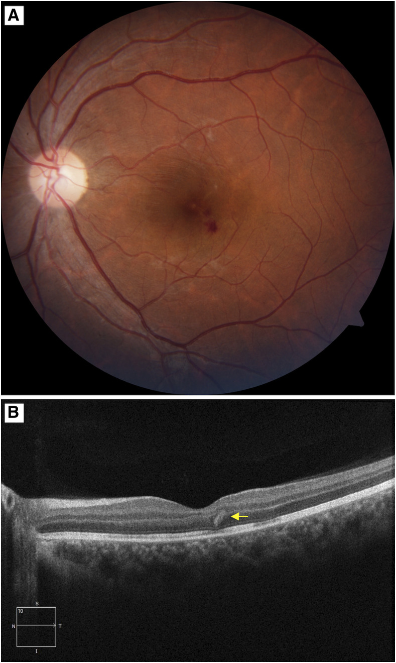
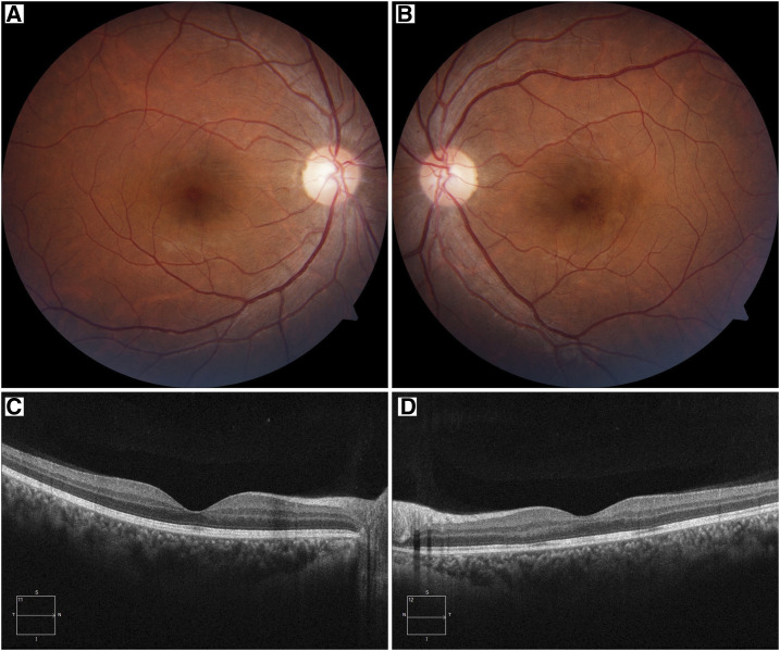
We describe a case of dengue fever-associated foveolitis that presented initially to the ophthalmologists with complaints of unilateral diminution of vision. A 30-year-old Indian woman had presented with sudden onset diminution of vision in the left eye (LE) for the past 2 days. It was also associated with low-grade fever and myalgia, which started few days before visual deterioration. Fundus showed few retinal hemorrhages and tiny subretinal yellowish lesions at the fovea in the LE. Optical coherence tomography and fluorescein angiography were indicative of foveolitis. Amsler charting showed a central scotoma in the LE. She was treated with oral steroids along with supportive treatments. A near-complete anatomical and functional recovery was noted. Our case depicts the significance of awareness of the ophthalmic complications of dengue fever among both ophthalmologists and physicians, and also highlights the key clinical and multimodal imaging findings in a case of dengue foveolitis.
Figures


References
-
- World Health Organization, Regional Office for South-East Asia , 2011. Comprehensive Guidelines for Prevention and Control of Dengue and Dengue Haemorrhagic Fever—Revised and Expanded Edition. Geneva, Switzerland: WHO; Available at: www.searo.who.int/entity/vector_borne_tropical_diseases/documents/SEAROT.... Accessed June 14, 2020.
-
- Teixeira MG, Barreto M, 2009. Diagnosis and management of dengue: clinical review. BMJ 339: 1189–1193. - PubMed
-
- Gupta P, Jain C, Aggarwal A, Gupta SC, 2011. Dengue fever presenting with macular hemorrhages. Retin Cases Brief Rep 5: 213–218. - PubMed
-
- Aggarwal A, Chandra J, Aneja S, Patwri AK, Dutta AK, 1998. An epidemic of dengue haemorrhagic fever and dengue shock syndrome in children in Delhi. Indian Pediatr 35: 727–732. - PubMed
Publication types
MeSH terms
Substances
LinkOut - more resources
Full Text Sources
Other Literature Sources
Medical
Miscellaneous
