. 2020 Dec;8(6):e00673.
doi: 10.1002/prp2.673.

Allosteric modulation of adenosine A1 and cannabinoid 1 receptor signaling by G-peptides

Affiliations

Allosteric modulation of adenosine A1 and cannabinoid 1 receptor signaling by G-peptides

Anja M Touma et al. Pharmacol Res Perspect. 2020 Dec.

Abstract

While allosteric modulation of GPCR signaling has gained prominence to address the need for receptor specificity, efforts have mainly focused on allosteric sites adjacent to the orthosteric ligand-binding pocket and lipophilic molecules that target transmembrane helices. In this study, we examined the allosteric influence of native peptides derived from the C-terminus of the Gα subunit (G-peptides) on signaling from two Gi-coupled receptors, adenosine A1 receptor (A1 R) and cannabinoid receptor 1 (CB1 ). We expressed A1 R and CB1 fusions with G-peptides derived from Gαs, Gαi, and Gαq in HEK 293 cells using systematic protein affinity strength modulation (SPASM) and monitored the impact on downstream signaling in the cell compared to a construct lacking G-peptides. We used agonists N6 -Cyclopentyladenosine (CPA) and 5'-N-Ethylcarboxamidoadenosine (NECA) for A1 R and 2-Arachidonoylglycerol (2-AG) and WIN 55,212-2 mesylate (WN) for CB1 . G-peptides derived from Gαi and Gαq enhance agonist-dependent cAMP inhibition, demonstrating their effect as positive allosteric modulators of Gi-coupled signaling. In contrast, both G-peptides suppress agonist-dependent IP1 levels suggesting that they differentially function as negative allosteric modulators of Gq-coupled signaling. Taken together with our previous studies on Gs-coupled receptors, this study provides an extended model for the allosteric effects of G-peptides on GPCR signaling, and highlights their potential as probe molecules to enhance receptor specificity.

Keywords: CB1; GTP-binding proteins; N(6)-cyclopentyladenosine; SCH 442 416; adenosine A1; adenosine-5’-(N-ethylcarboxamide); cannabinoid; receptor; signal transduction.


Figures

FIGURE 1
FIGURE 1
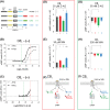
Gα peptides differentially impact Gs and Gi signaling in Cannabinoid (CB1) receptors. A, SPASM sensors for characterization of second messenger response. Schematics of the A1R and CB1 GPCR peptide sensors containing C‐terminal Gα peptides corresponding to s‐, i‐, or q‐ 5α helices separated with Gly‐Ser‐Gly (GSG)4 linkers to ensure rotational freedom. The no‐pep (−) construct lacks the Gα C‐terminal peptide. Forskolin‐stimulated cAMP dose‐response curves of B, CB1 agonist, 2‐Arachidonoylglycerol (2‐AG), and C, WIN 55,212‐2 mesylate (WN55212‐2) in a CB1 no‐pep (−) sensor (representative curves from N = 2 independent biological replicates composed of ≥3 technical repeats each). cAMP levels shown in the absence (gray line) and presence (black line) of pertussis toxin (PTX) treatment. Ligands potentiate forskolin‐stimulated cAMP accumulation at 30 μmol/L, suggesting Gs bias (B and C, red dashed lines). 2‐AG and WIN 55,212‐2 mesylate (WN) inhibit forskolin‐stimulated cAMP at 50 nmol/L and 300 nmol/L, respectively, suggesting Gi bias (B and C, green dashed lines). cAMP levels of tethered CB1 sensors after stimulation by forskolin and 30 μmol/L 2‐AG (D) or WN (E) (N = 5 independent biological replicates). F, summary of Gα peptide influence on Gs signaling and cAMP production in CB1. Inhibition of forskolin‐stimulated cAMP by tethered CB1 sensors after stimulation by 50 nmol/L 2‐AG (G) (N = 8 independent biological replicates) or 300 nmol/L WN (H) (N = 6 independent biological replicates). I, summary of Gα peptide influence on Gi signaling and cAMP inhibition in CB1. GPCR‐Gα C‐terminal peptide sensors are compared with the no‐pep (−) control. Results are expressed as mean ± SE. ****P < .0001; *P < .05
FIGURE 2
FIGURE 2
Characterization of cAMP modulation in adenosine receptor (A1R) by SPASM sensors. Forskolin‐stimulated cAMP dose‐response curves of (A), A1R agonist, N6‐Cyclopentyladenosine (CPA). cAMP levels shown in the absence (gray line) and presence (black line) of pertussis toxin (PTX) treatment. 50 nmol/L CPA inhibits forskolin‐stimulated cAMP, suggesting Gi bias (A, green dashed line). B, Inhibition of forskolin‐stimulated cAMP by tethered A1R peptide sensors after stimulation by 50 nmol/L CPA. C, Summary of Gα peptide influence on Gi signaling and cAMP inhibition. GPCR‐Gα C‐terminal peptide sensors are compared with the no‐pep (−) control. Results are expressed as mean ± SE. ***P < .001; **P < .01. N = 8 independent biological replicates
FIGURE 3
FIGURE 3
Gαq and Gαi peptides inhibit signaling through Gq. A, IP1 dose‐response curve of A1R agonists, CPA (gray lines) (representative curves from N = 2 independent biological replicates composed of ≥3 technical repeats each) and NECA (black line) (N = 3 technical repeats), with A1R‐no‐pep (−) sensor. IP1 levels shown in the absence (dark gray line) and presence (light gray line) of pertussis toxin (PTX) treatment. 100 μmol/L CPA or NECA stimulate IP1 (A, blue dashed line). B, IP1 signal from A1R after stimulation by 100 μmol/L CPA (left) (N = 3 independent biological replicates) or NECA (right) (N = 4 independent biological replicates) in the presence of different Gα C‐terminal peptides compared to no‐pep (−) control. c, summary of Gα peptide influence on Gq signaling and IP1 production in A1R. D, IP1 dose‐response curve of CB1 agonists, 2‐AG (gray lines) (N = 5 independent biological replicates) and WN (black line) (N = 4 independent biological replicates), with CB1‐no‐pep (−) sensor. IP1 levels shown in the absence (dark gray line) and presence (light gray line) of pertussis toxin (PTX) treatment. 100 μmol/L 2‐AG or WN stimulate IP1 (D, blue dashed line). E, IP1 signal from CB1 Gα C‐terminal peptide sensors after stimulation by 100 μmol/L 2‐AG (left) or WN (right) compared to no‐pep (−) control (N = 3 technical repeats). F, summary of Gα peptide influence on Gq signaling and IP1 production in CB1. Results are expressed as mean ± SE. ****P < .0001; ***P < .001; **P < .01

References

    1. Insel PA, Tang C‐M, Hahntow I, Michel MC. Impact of GPCRs in clinical medicine: monogenic diseases, genetic variants and drug targets. Biochim Biophys Acta. 2007;1768:994‐1005. 10.1016/j.bbamem.2006.09.029 - DOI - PMC - PubMed
    1. Conn PJ, Christopoulos A, Lindsley CW. Allosteric modulators of GPCRs: a novel approach for the treatment of CNS disorders. Nat Rev Drug Discov. 2009;8:41‐54. 10.1038/nrd2760 - DOI - PMC - PubMed
    1. Foster DJ, Conn PJ. Allosteric modulation of GPCRs: new insights and potential utility for treatment of schizophrenia and other CNS disorders. Neuron. 2017;94:431‐446. 10.1016/j.neuron.2017.03.016 - DOI - PMC - PubMed
    1. Chung KY. Structural aspects of GPCR‐G protein coupling. Toxicol Res. 2013;29:149‐155. 10.5487/TR.2013.29.3.149 - DOI - PMC - PubMed
    1. Wakefield AE, Mason JS, Vajda S, Keserű GM. Analysis of tractable allosteric sites in G protein‐coupled receptors. Sci Rep. 2019;9:6180 10.1038/s41598-019-42618-8 - DOI - PMC - PubMed

Publication types

MeSH terms

Substances

LinkOut - more resources