. 2021 May 7;13(2):104-115.
doi: 10.1093/jmcb/mjaa059.

Early development and functional properties of tryptase/chymase double-positive mast cells from human pluripotent stem cells

Affiliations

Early development and functional properties of tryptase/chymase double-positive mast cells from human pluripotent stem cells

Guohui Bian et al. J Mol Cell Biol. .

Abstract

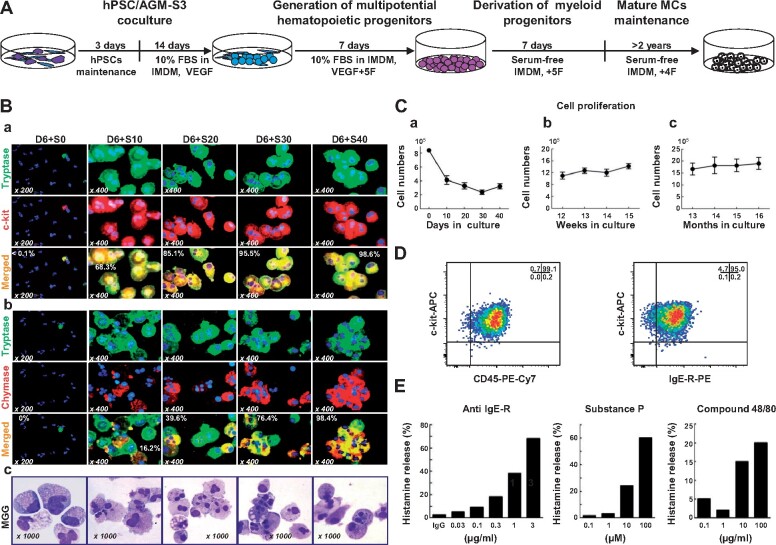
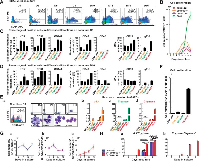
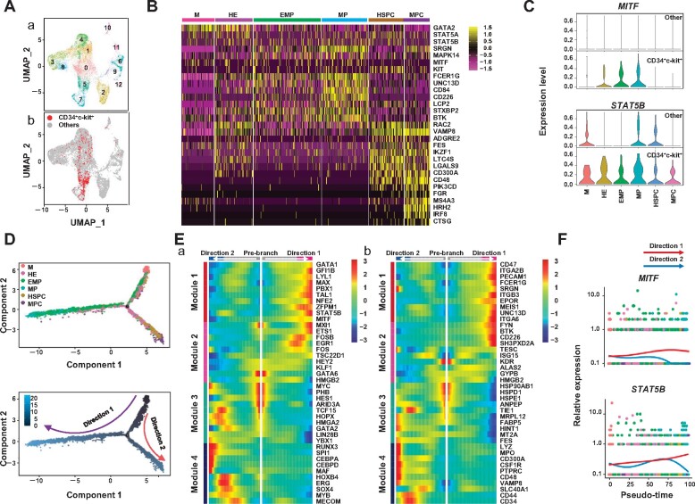
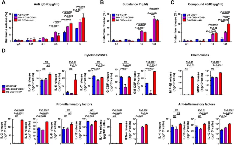
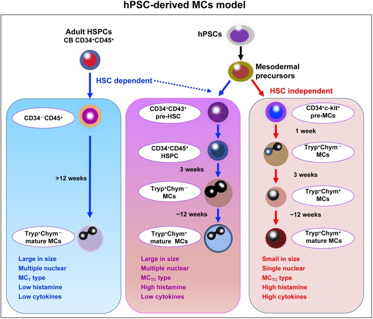
Mast cells (MCs) play a pivotal role in the hypersensitivity reaction by regulating the innate and adaptive immune responses. Humans have two types of MCs. The first type, termed MCTC, is found in the skin and other connective tissues and expresses both tryptase and chymase, while the second, termed MCT, which only expresses tryptase, is found primarily in the mucosa. MCs induced from human adult-type CD34+ cells are reported to be of the MCT type, but the development of MCs during embryonic/fetal stages is largely unknown. Using an efficient coculture system, we identified that a CD34+c-kit+ cell population, which appeared prior to the emergence of CD34+CD45+ hematopoietic stem and progenitor cells (HSPCs), stimulated robust production of pure Tryptase+Chymase+ MCs (MCTCs). Single-cell analysis revealed dual development directions of CD34+c-kit+ progenitors, with one lineage developing into erythro-myeloid progenitors (EMP) and the other lineage developing into HSPC. Interestingly, MCTCs derived from early CD34+c-kit+ cells exhibited strong histamine release and immune response functions. Particularly, robust release of IL-17 suggested that these early developing tissue-type MCTCs could play a central role in tumor immunity. These findings could help elucidate the mechanisms controlling early development of MCTCs and have significant therapeutic implications.

Keywords: chymase; development; human pluripotent stem cells (hPSCs); mast cells; tryptase.


Figures

Figure 1
Figure 1
Early hPSC-MCs share skin MC protease phenotypes and functions. (A) Schematic illustrating the strategy for the generation of MCs from D14 hPSC/AGM-S3 coculture. (B) Immunostaining and MGG staining of early developed hPSC-MCs over the time course of SF-MC cultivation. (C) Persistence of cell proliferation of H1/AGM-S3 coculture D14 (a), short-term (up to 15 weeks, b), and long-term (up to 16 months, c) in SF-MC cultures. (D) Mature hPSC-MCs (20 weeks in SF-MC culture) co-expressing CD45, c-kit, and a high-affinity IgE-R. (E) Histamine release by mature hPSC-MCs following stimulation with an antibody against the human IgE-R (CRA1), substance P, and compound 48/80, respectively.
Figure 2
Figure 2
Rapid maturation of MCTCs generated from a CD34+c-kit+ cell population. (A) Flow cytometric profiles showing expression of CD34+c-kit+ cells derived from H1/AGM-S3 coculture. (B) Proliferation of CD34+CD45+, CD34+CD43+, CD34+c-kit+, and CD45+ cell fractions on D4‒D16 derived from 1 × 104 H1 cells (independent experiments, n =3; mean ± SD). (C and D) Flow cytometric analysis showing representative phenotypic expression of mesoderm (KDR), endothelial (CD31 and CD144), hematopoietic (CD43 and CD45), and MC (CD13 and IgE-R) markers in the four cell fractions at D8 (C) and D16 (D) during a single culture. (E) Four cell fractions defined by expression of CD34 and c-kit were sorted by FACS from a D8 H1/AGM-S3 coculture. MGG analysis showing typical morphologies of the sorted CD34+c-kit+, CD34+c-kit, CD34c-kit+, and CD34c-kit cell fractions (a) and qPCR analysis showing mRNA expression of c-kit (b), tryptase (c), and chymase (d) in each cell fraction, total coculture D8 cells, and a pure population of MCs. Independent experiments, n =3; mean ± SD. ND, not detected. Scale bar, 10 µm. (F) Total cell proliferation of the four cell fractions sorted from D8. (G) Total cell proliferation of CB CD34+ cells (a), coculture D14 CD34+CD45+ cells (b), and coculture D8 CD34+c-kit+ cells (c) in SF-MC culture. (H) Compared with CB CD34+ and coculture D14 CD34+CD45+ progenitor cells, a high proportion (36.3% ± 3.4%) of coculture D8 CD34+c-kit+ cell-derived c-kit+Tryptase+ MCs could be observed early on D7 in SF-MC culture and reached a purity of 90.3% ± 1.4% after 21 days in culture (a) that was associated with a rapid increase in co-expression of chymase (90.2% ± 1.1% at D21, b). NS, non-significant.
Figure 3
Figure 3
Dual development potential of MCs before and after HSPCs appeared. (A) Identification of unsupervised clustering for 2 days combined with UMPA. (a) Each dot represents one cell, and colors represent cell clusters as indicated. (b) Expression of CD34+c-kit+ cells in different clusters. (B) Heatmap showing scaled expression of MC-related genes in the six clusters. (C) Violin plots showing expression of featured genes in each cell cluster between CD34+c-kit+ cells and other cells. (D) Trajectory analysis by Monocle 2 combining six clusters from D5 with 13 clusters from D5_5 indicates the development of CD34+c-kit+ cells. (E) Four distinct gene expression patterns along the pseudotime axis were inferred by Monocle 2. Prominent transcription factors (a) and other genes (b) are listed. (F) Expression of representative genes of pattern 1 along pseudotime axis inferred by Monocle 2.
Figure 4
Figure 4
Histamine and cytokine release by mature MCs derived from three different protocols. (AC) IgE-sensitized MCs were stimulated for 30 min with anti-IgE-R (CRA1), Substance P, and compound 48/80. Data are expressed as mean ± SD of at least three sample replicates. (D) Heterogenous cytokine release after anti-IgE-R stimulation. Cytokine levels are expressed as pg/106 cells. Significant differences among MC culture protocols are indicated. Data are expressed as mean ± SD of at least three sample replicates. Blue, CB CD34+-MCs; purple, coculture D14 CD34+CD45+-MCs; red, coculture D8 CD34+c-kit+-MCs. NS, non-significant.
Figure 5
Figure 5
Model depicting the development pathway for hPSC-MCs. There is a specific development stage during MCTCs from hPSC/AGM-S3 coculture. Early MCTCs are derived from CD34+c-kit+ cells, independent of HSCs. Compared with the MCs derived from HSC-dependent pathway, these cells exhibit special morphology and function.

References

    1. Abrink M., Grujic M., Pejler G. (2004). Serglycin is essential for maturation of mast cell secretory granule. J. Biol. Chem. 279, 40897–40905. - PubMed
    1. Álvarez-Errico D., Oliver-Vila I., Ainsua-Enrich E., et al. (2011). CD84 negatively regulates IgE high-affinity receptor signaling in human mast cells. J. Immunol. 187, 5577–5586. - PMC - PubMed
    1. Bachelet I., Munitz A., Mankutad D., et al. (2006). Mast cell costimulation by CD226/CD112 (DNAM-1/Nectin-2): a novel interface in the allergic process. J. Biol. Chem. 281, 27190–27196. - PubMed
    1. Bin N.R., Jung C.H., Piggott C., et al. (2013). Crucial role of the hydrophobic pocket region of Munc18 protein in mast cell degranulation. Proc. Natl Acad. Sci. USA 110, 4610–4615. - PMC - PubMed
    1. Butler A., Hoffman P., Smibert P., et al. (2018). Integrating single-cell transcriptomic data across different conditions, technologies, and species. Nat. Biotechnol. 36, 411–420. - PMC - PubMed

Publication types

MeSH terms