. 2020 Oct 30;18(10):e3000958.
doi: 10.1371/journal.pbio.3000958. eCollection 2020 Oct.

Differences in structure and hibernation mechanism highlight diversification of the microsporidian ribosome

Affiliations

Differences in structure and hibernation mechanism highlight diversification of the microsporidian ribosome

Kai Ehrenbolger et al. PLoS Biol. .

Abstract

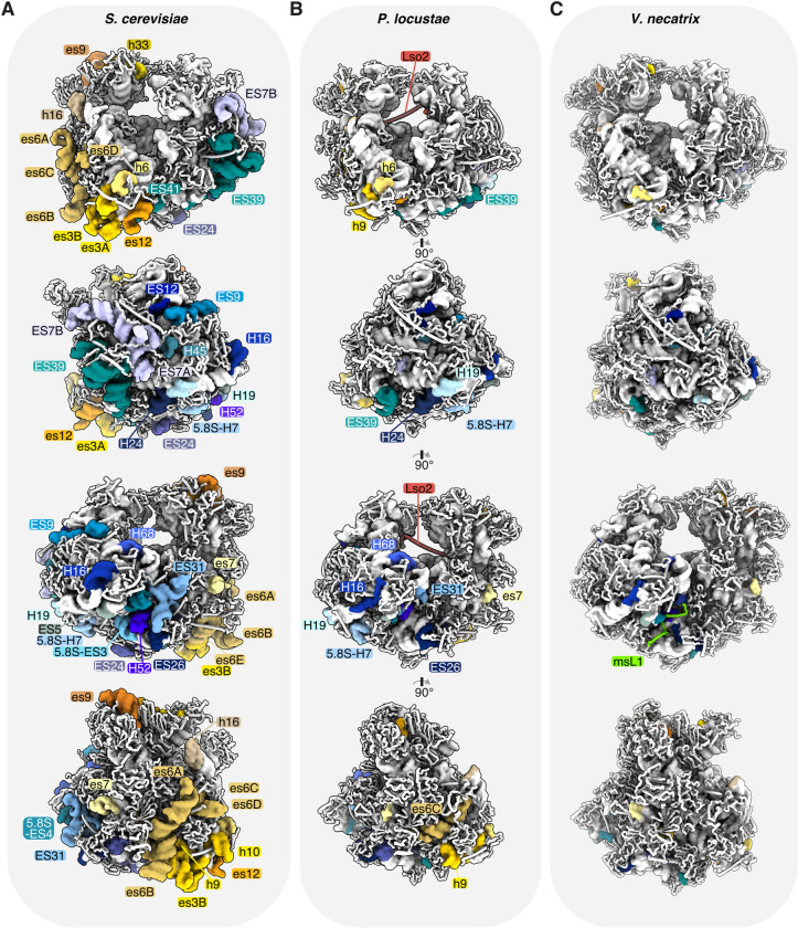
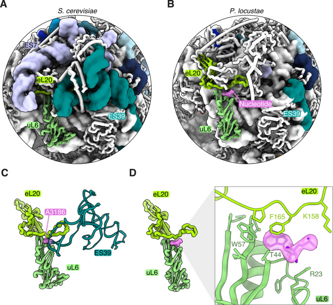
Assembling and powering ribosomes are energy-intensive processes requiring fine-tuned cellular control mechanisms. In organisms operating under strict nutrient limitations, such as pathogenic microsporidia, conservation of energy via ribosomal hibernation and recycling is critical. The mechanisms by which hibernation is achieved in microsporidia, however, remain poorly understood. Here, we present the cryo-electron microscopy structure of the ribosome from Paranosema locustae spores, bound by the conserved eukaryotic hibernation and recycling factor Lso2. The microsporidian Lso2 homolog adopts a V-shaped conformation to bridge the mRNA decoding site and the large subunit tRNA binding sites, providing a reversible ribosome inactivation mechanism. Although microsporidian ribosomes are highly compacted, the P. locustae ribosome retains several rRNA segments absent in other microsporidia, and represents an intermediate state of rRNA reduction. In one case, the near complete reduction of an expansion segment has resulted in a single bound nucleotide, which may act as an architectural co-factor to stabilize a protein-protein interface. The presented structure highlights the reductive evolution in these emerging pathogens and sheds light on a conserved mechanism for eukaryotic ribosome hibernation.


Conflict of interest statement

The authors have declared that no competing interests exist.

Figures

Fig 1
Fig 1. The cryo-EM structure of the P. locustae ribosome, bound to Lso2 and a structural nucleotide.
Composite cryo-EM map consisting of maps focused on the LSU, SSU-body, and SSU-head is shown (EMD-11437). The complete ribosome is shown in the center, while the SSU (left) and LSU (right) are depicted in isolation on both sides. The SSU is colored in shades of yellow (RNA in gold, proteins in light yellow), while the LSU is colored in shades of blue (RNA in dark blue, proteins in light blue), with selected ribosomal proteins labeled and colored in shades of green. The hibernation and recovery factor Lso2 is highlighted in red. The inset showcases the nucleotide-binding site (purple) at the interface between the 2 LSU proteins uL6 and eL20 (shades of green), displayed by superimposing the cryo-EM map with the molecular model. C, C-terminus; cryo-EM, cryo–electron microscopy; E-tRNA, exit site tRNA; LSU, large subunit; N, N-terminus; SSU, small subunit.
Fig 2
Fig 2. Microsporidian Lso2 interactions with the ribosome.
(A) Slab view of Lso2 (red) bound ribosomes along with the cryo-EM density for E-site tRNA (sky blue). Lso2 ends contacting the SSU and LSU are indicated as N and C, respectively (PDB 6ZU5). (B) Lso2 prevents tRNA and mRNA binding in the A- and P- site as shown by the superimposed tRNAs (aquamarine, from PDB 4V6F) and an mRNA (pink surface, from PDB 4V6F). (C) An isolated view of Lso2 sterically blocking the codon–anticodon site, as well as the D- and T-arm of the P-site tRNA. The C-terminal end overlaps with the T-arm of the A-site tRNA. (D–F) Molecular contacts between Lso2 and the ribosome, shown as cryo-EM density (mesh) and the structural model. Lso2 residues contacting the rRNA or ribosomal proteins are indicated. A-site, acceptor site; A-tRNA, acceptor site tRNA; C, C-terminus; cryo-EM, cryo–electron microscopy; E-site; exit site; E-tRNA, exit site tRNA; LSU, large subunit; N, N-terminus; P-site, peptidyl site; P-tRNA, peptidyl site tRNA; SSU, small subunit.
Fig 3
Fig 3. Ribosomal RNA compaction in microsporidia.
Comparative analysis of expansion segments and the absence thereof between (A) S. cerevisiae (PDB 4V88), (B) P. locustae (PDB 6ZU5), and (C) V. necatrix (PDB 6RM3). (A–C) Four 90°-related views of the ribosome are shown (from top to bottom), with expansion segments colored and labeled in shades of yellow and orange (small subunit) or shades of blue and green (large subunit).
Fig 4
Fig 4. Reductive evolution of ES39 to a single structural nucleotide.
A comparison of ES7 and ES39 between (A) S. cerevisiae (PDB 4V88) and (B) P. locustae (PDB 6ZU5). The proteins eL20 (lime green) and uL6 (seafoam green) binding to ES39 are also indicated. (C) An isolated, close-up view of the binding interface between eL20 and uL6, stabilized by A3186 (pink) from ES39 in the S. cerevisiae ribosome (PDB 4V88). (D) A single nucleotide (pink) is bound to the protein–protein interface in P. locustae. The inset depicts the cryo–electron microscopy map consistent with a single nucleotide between eL20 and uL6.

References

    1. Maitra A, Dill KA. Bacterial growth laws reflect the evolutionary importance of energy efficiency. Proc Natl Acad Sci U S A. 2015;112:406–11. 10.1073/pnas.1421138111 - DOI - PMC - PubMed
    1. Tempest DW, Neijssel OM. The status of YATP and maintenance energy as biologically interpretable phenomena. Annu Rev Microbiol. 1984;38:459–513. 10.1146/annurev.mi.38.100184.002331 - DOI - PubMed
    1. Valcourt JR, Lemons JMS, Haley EM, Kojima M, Demuren OO, Coller HA. Staying alive: metabolic adaptations to quiescence. Cell Cycle. 2012;11:1680–96. 10.4161/cc.19879 - DOI - PMC - PubMed
    1. Prossliner T, Skovbo Winther K, Sørensen MA, Gerdes K. Ribosome hibernation. Annu Rev Genet. 2018;52:321–48. 10.1146/annurev-genet-120215-035130 - DOI - PubMed
    1. Sharma MR, Dönhöfer A, Barat C, Marquez V, Datta PP, Fucini P, et al. PSRP1 is not a ribosomal protein, but a ribosome-binding factor that is recycled by the Ribosome-recycling Factor (RRF) and Elongation Factor G (EF-G). J Biol Chem. 2010;285:4006–14. 10.1074/jbc.M109.062299 - DOI - PMC - PubMed

Publication types