Review
. 2021 Jan 28;42(1):29-55.
doi: 10.1210/endrev/bnaa025.

Microvascular Dysfunction in Diabetes Mellitus and Cardiometabolic Disease

Affiliations
Review

Microvascular Dysfunction in Diabetes Mellitus and Cardiometabolic Disease

William B Horton et al. Endocr Rev. .

Abstract

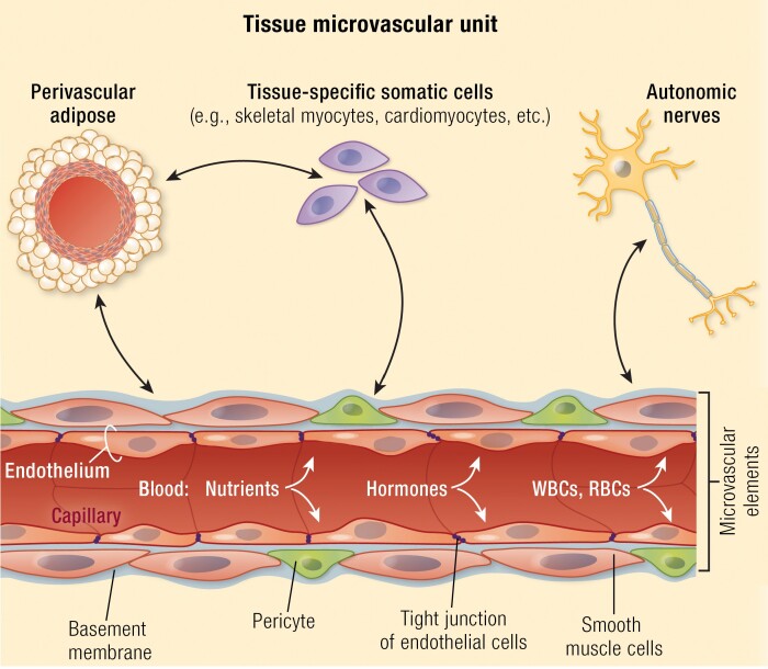
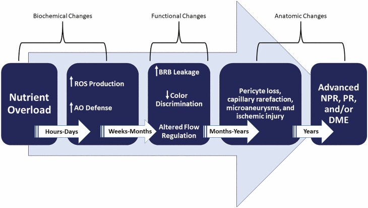
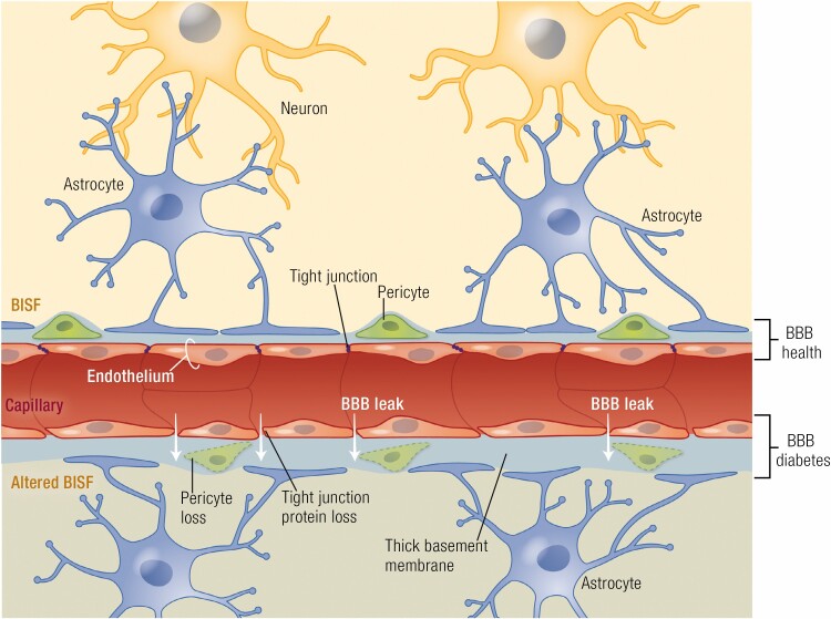
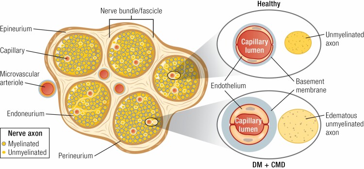
This review takes an inclusive approach to microvascular dysfunction in diabetes mellitus and cardiometabolic disease. In virtually every organ, dynamic interactions between the microvasculature and resident tissue elements normally modulate vascular and tissue function in a homeostatic fashion. This regulation is disordered by diabetes mellitus, by hypertension, by obesity, and by dyslipidemia individually (or combined in cardiometabolic disease), with dysfunction serving as an early marker of change. In particular, we suggest that the familiar retinal, renal, and neural complications of diabetes mellitus are late-stage manifestations of microvascular injury that begins years earlier and is often abetted by other cardiometabolic disease elements (eg, hypertension, obesity, dyslipidemia). We focus on evidence that microvascular dysfunction precedes anatomic microvascular disease in these organs as well as in heart, muscle, and brain. We suggest that early on, diabetes mellitus and/or cardiometabolic disease can each cause reversible microvascular injury with accompanying dysfunction, which in time may or may not become irreversible and anatomically identifiable disease (eg, vascular basement membrane thickening, capillary rarefaction, pericyte loss, etc.). Consequences can include the familiar vision loss, renal insufficiency, and neuropathy, but also heart failure, sarcopenia, cognitive impairment, and escalating metabolic dysfunction. Our understanding of normal microvascular function and early dysfunction is rapidly evolving, aided by innovative genetic and imaging tools. This is leading, in tissues like the retina, to testing novel preventive interventions at early, reversible stages of microvascular injury. Great hope lies in the possibility that some of these interventions may develop into effective therapies.

Keywords: cardiometabolic disease; diabetes complications; diabetes mellitus; microvascular rarefaction; microvessels; vascular endothelium.


Figures

Graphical Abstract
Graphical Abstract
In each of the 6 tissues reviewed, we highlight the reciprocal relationship(s) between the tissue’s somatic cells and the microvasculature serving them. Together these components function as a microvascular unit. Early in their course, diabetes mellitus and cardiometabolic disease disrupt these microvascular units and produce tissue dysfunction. Over time, this disruption leads to common (eg, increased endothelial barrier permeability, pericyte loss, capillary rarefaction, disordered angiogenesis, etc.) as well as tissue-specific (eg, microglia activation in the central nervous system and retina, sympathetic overactivity, and perivascular adipose inflammation in peripheral tissues) microvascular injury responses that are orchestrated by a host of both systemic and local signaling processes.
Figure 1.
Figure 1.
An approximate time course for the development of microvascular lesions within the retina neurovascular unit. Early biochemical effects from hyperglycemia (or deficient insulin action) are marked by increased production of ROS (eg, superoxide, peroxynitrate, hydrogen peroxide, etc.). Less well defined, but apparent in other tissues, is activation of antioxidant defense mechanisms. With time, functional changes in the neurovascular unit of the retina occur with increased permeability of capillaries as well as changes in the neural retina that affect contrast and color discrimination and alter blood flow regulation. These typically antedate early anatomic changes that include pericyte loss, capillary dropout, micro-aneurysms, venous beading, and intraretinal microvascular abnormalities. With longer duration of hyperglycemia, more advanced forms of retinopathy (eg, NPR and PR) as well as DME develop. ROS, reactive oxygen species; AO, antioxidant; BRB, blood–retinal barrier; NPR, nonproliferative retinopathy; PR, proliferative retinopathy; DME, diabetic macular edema.
Figure 2.
Figure 2.
The brain NVU (schematized here at the capillary level) includes neurons which reciprocally signal via astrocytes to the microvasculature of small arterioles and capillaries. This signaling affects tone of SMCs and pericytes and regulates EC permeability properties. The astrocyte foot processes also limit passage of macromolecules to BISF. In health, the low permeability of the blood-brain barrier depends critically on the tight junctions of ECs but also requires normal function of pericytes, astrocytes, and the basement membrane. With long-standing DM (lower portion of figure) there is increased BBB permeability due to loss of tight junction proteins. Pericytes, which cover much of the capillary vasculature, are lost facilitating the permeability increases. Over time, ECs are also lost and capillary rarefaction occurs, thus altering the composition of BISF. NVU, neurovascular unit; BBB, blood–brain barrier; DM, diabetes mellitus; SMC, smooth muscle cells; EC, endothelial cell; BISF, brain interstitial fluid.
Figure 3.
Figure 3.
Unmyelinated and myelinated peripheral nerve bundles are ensconced in the outer layer of connective tissue (epineurium) and an inner band (perineurium) that delineates the nerve fascicles. Microvascular arterioles penetrate both layers bringing a capillary supply to the endoneurium (a peripheral nerve analog of cerebrospinal fluid). In health, the capillary endothelium maintains a tight barrier regulating glucose, oxygen, and electrolyte access to axonal elements in the endoneurium. With DM and CMD, the capillary basement membrane expands and there is endothelial hypertrophy and increased permeability resulting in diminished flow and oxygen delivery, axon edema, and subsequent nerve dysfunction. Injury induced by hyperglycemia is exacerbated by other CMD factors (eg, hypertension, obesity, dyslipidemia, etc.). Chronic capillary rarefaction produces worsened ischemia and axon degeneration. DM, diabetes mellitus; CMD, cardiometabolic disease.
Figure 4.
Figure 4.
In health, inflowing blood through the feed artery is progressively distributed to smaller and smaller arterioles within the muscle microvasculature, ending in a capillary network fed by terminal arterioles paired with draining venules. To facilitate blood flow distribution that matches their oxygen and nutrient needs, myocytes release vasoactive substances into the draining venules (green arrows) with signaling transmitted to adjacent segments of terminal arterioles. The latter can signal both proximally and distally (black 2-headed arrows) via gap junctions abundant in the vascular endothelium, thereby increasing the volume of vasculature dilated (or constricted) in response to the myocytes’ needs. Overall flow is regulated by myogenic autoregulation, by sympathetic alpha-adrenergic tone, by signals from the myocytes themselves, and by circulating hormones (eg, insulin). Obesity, hypertension, insulin resistance, and DM can chronically decrease muscle perfusion by actions at the feed artery and at first and second arterioles (in part through increased sympathetic tone). More distally, these CMD disorders also impair the ability of insulin to perfuse capillary beds, depriving them of nutrients and oxygen. DM, diabetes mellitus; CMD, cardiometabolic disease.
Figure 5.
Figure 5.
CPVAT is located adjacent to various macro- and microvascular surfaces, including coronary arterial media and adventitial vasa vasorum, which enables paracrine secretion of many vasoactive substances. In pathophysiological states (eg, DM and CMD), CPVAT paracrine secretion becomes unbalanced and dysfunctional, resulting in reduced NO bioavailability and enhanced secretion of pro-inflammatory adipokines that increase large artery stiffness and contribute to downstream microvascular (ie, vasa vasorum) dysfunction. PVAT, perivascular adipose tissue; DM, diabetes mellitus; CMD, cardiometabolic disease; NO, nitric oxide; ET-1, endothelin-1; TNFα, tumor necrosis factor alpha).
Figure 6.
Figure 6.
Studies of the renal microvascular effects of diabetes mellitus and cardiometabolic disease have long focused on glomerular injury and control of glomerular perfusion by regulation of afferent and efferent arteriolar tone. In particular, blocking angiotensin II-induced efferent arteriole constriction and reducing glomerular capillary pressure has been a therapeutic mainstay. The beneficial effects of SGLT-2 inhibition on renal function may relate in part to increased sodium delivery to the macula densa of the distal tubule, with resulting decreased renin production and angiotensin II generation. Further renal benefit may derive from (1) decreased energy expenditure for sodium reabsorption in the proximal tubule, lessening oxygen consumption from peritubular postglomerular capillaries and diminishing renal cortical hypoxic stress, epithelial cell apoptosis and tubular fibrosis, and (2) from increased adenosine release at the macula densa which acts to vasoconstrict the afferent and vasodilate the efferent arteriole. Ang II, angiotensin II; SGLT2i, sodium-glucose cotransporter-2 inhibition.

Similar articles

Cited by

References

    1. Segal SS, Jacobs TL. Role for endothelial cell conduction in ascending vasodilatation and exercise hyperaemia in hamster skeletal muscle. J Physiol. 2001;536(Pt 3):937-946. - PMC - PubMed
    1. Honig CR, Odoroff CL, Frierson JL. Capillary recruitment in exercise: rate, extent, uniformity, and relation to blood flow. Am J Physiol. 1980;238(1):H31-H42. - PubMed
    1. Segal SS. Regulation of blood flow in the microcirculation. Microcirculation. 2005;12(1):33-45. - PubMed
    1. Hill J, Rom S, Ramirez SH, Persidsky Y. Emerging roles of pericytes in the regulation of the neurovascular unit in health and disease. J Neuroimmune Pharmacol. 2014;9(5):591-605. - PMC - PubMed
    1. Gardner TW, Davila JR. The neurovascular unit and the pathophysiologic basis of diabetic retinopathy. Graefes Arch Clin Exp Ophthalmol. 2017;255(1):1-6. - PMC - PubMed

Publication types