Review
. 2020 Oct 30;10(1):27.
doi: 10.1186/s12348-020-00217-z.

Susac syndrome (Retino-cochleo-cerebral vasculitis), the ophthalmologist in the role of the whistleblower

Affiliations
Review

Susac syndrome (Retino-cochleo-cerebral vasculitis), the ophthalmologist in the role of the whistleblower

Ioannis Papasavvas et al. J Ophthalmic Inflamm Infect. .

Abstract

Background/purpose: Susac syndrome is a rare microangiopathy of suspected autoimmune origin affecting arteries of the retina, the cochlea and the brain. The aim of the study was to give a review of the disease entity and determine the proportion of cases and their characteristics in a uveitis referral centre.

Patients and methods: Charts of patients with the diagnosis of Susac syndrome seen in the Uveitis Clinic of the Centre for Ophthalmic Specialised Care (COS), Lausanne, Switzerland were reviewed retrospectively to determine the frequency of such cases in a uveitis referral centre. Clinical symptoms and signs, functional data, imaging signs and evolution were analysed in the 3 COS cases and one case shared with the Uveitis Clinic of the Department of Ophthalmology, University of Innsbruck, Austria. Characteristic signs were searched possibly allowing a prompt diagnosis.

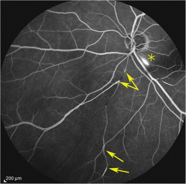
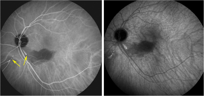
Results: During the period from 1994 to 2019 (24 years, 2045 patients), 3 charts with the diagnosis of Susac syndrome were found (0.15%). The whole collective, including the additional case, comprised three women aged 28, 32 and 63 at presentation and one man, aged 42. None of the 3 cases that were referred were diagnosed beforehand. The characteristic item found in all 4 cases was the abrupt arterial stop or segmental interruption of arteries and increased staining of arterial wall on angiography more clearly shown on indocyanine green angiography that can potentially be proposed as a crucial diagnostic element. All 4 cases responded to dual steroidal and non-steroidal immunosuppression. Under treatment, all four patients did not show any further evolution.

Conclusion: Susac syndrome is a multilocation arteritis of the head that can involve the eye, ear and brain often first diagnosed by the ophthalmologist. The diagnosis is rapidly reached in uveitis referral centres but seems to be missed otherwise, A helpful angiographic sign to be searched is an abrupt or segmental arterial stop and increased staining of the arterial wall more clearly seen on indocyanine green angiography. Patients often present first to the ophthalmologist who should be acting as a whistleblower to avoid severe involvement of the brain.

Keywords: Branch retinal artery occlusion (BRAO); Fluorescein angiography; Indocyanine green angiography; Susac syndrome.


Conflict of interest statement

There is no financial disclosure.

Figures

Fig. 1
Fig. 1
Six weeks after presentation the ischaemic zone resulted in atrophy the of the inner retina; shown by OCT imaging (arrow); note that photoreceptor outer segment ellipsoid zone is conserved (case 1)
Fig. 2
Fig. 2
White-yellow discoloration of the course of a branch artery from inferior arcade (arrows) in a case evolving since 4 months (case 3)
Fig. 3
Fig. 3
Optical Coherence Tomography (OCT) shows oedematous retinal thickening in the infarcted area in a patient seen in the acute phase (case 1) The SLO fundus image clearly delineates the ischaemic area
Fig. 4
Fig. 4
a OCT imaging of the retina in a patient 4 months after retinal infarct showing atrophic thinning of the inner retina with conservation of the photoreceptor outer segments ellipsoid zone; note white course of involved artery on the SLO fundus picture (case 3). b Microperimetry clearly delineating the non-functional atrophic retinal area (case 3)
Fig. 5
Fig. 5
Fluorescein angiography (FA) showing abrupt interruption of two arteries inferiorly (arrows) and characteristic arterial wall hyperfluorescence (asterisk) (case 1)
Fig. 6
Fig. 6
Indocyanine green angiography. Same view of fundus as on Fig. 5, showing precisely arterial stop and/or interruption (arrows) (case 1)
Fig. 7
Fig. 7
Fluorescein angiography (FA) showing segmental artery occlusion and hyperfluorescence of the arterial walls in the left eye (two right pictures) in early (top) and late (bottom) angiographic phases. In the right eye (two left pictures) an area of non-perfusion (capillary drop) of vessels in the mid-periphery (top) with staining of arteries better seen in the late phase (bottom). (Case 2)
Fig. 8
Fig. 8
Vast upper scotoma corresponding to the ischaemic infarcted lower retina delineated by microperimetry (Fig. 9) (case 1)
Fig. 9
Fig. 9
Microperimetry showing the area of functional impairment caused by the retinal infarction. (Case 1)
Fig. 10
Fig. 10
Yellow-white zone of retinal infarction (case 1)
Fig. 11
Fig. 11
Panorama and posterior pole FA views showing arteritis with segmental involvement and abrupt artery occlusion or subocclusion (top sextet of frames). Posterior pole FA and ICGA views showing hyperfluorescence of the arteries on both FA (top two frames) and ICGA (bottom two frames) (arrows) and hypofluorescence in the areas of retinal infarction (asterisk) (bottom quartet of frames) (case 1)
Fig. 12
Fig. 12
Evolution of OCT views of infarcted zone in the acute phase (top) showing retinal oedema (arrow) and 6 months later (bottom) showing atrophy of inner retina (arrow) while external retina is conserved. (case 1)
Fig. 13
Fig. 13
Follow-up FA (top four frames) and ICGA bottom twoframes 5 weeks after acute episode and systemic corticosteroid treatment; complete resolution of arteritis (case 1)
Fig. 14
Fig. 14
Fundus picture OS taken 5 weeks after the acute episode showing quasi disappearance of whitish aspect in the infarcted zone (case 1)
Fig. 15
Fig. 15
Visual field and microperimetry 6 months after the acute episode, showing improved microperimetry score (left) and slight reduction of scotoma (right) (case 1)

References

    1. Papo T, Biousse V, LeHoang P, et al. Susac syndrome. Medicine (Baltimore) 77:3–11 - PubMed
    1. Susac JO, Hardman JM, Selhorst JB. Microangiopathy of the brain and retina. Neuroloogy 1979; 29: 313–316 - PubMed
    1. Susac JO. Susac’s syndrome: the triad of microangiopathy of the brain and retina with hearing loss in young women. Neurology. 1994;44:591–593. doi: 10.1212/WNL.44.4.591. - DOI - PubMed
    1. Dörr J, Krautwald S, Wildemann B, et al. Characteristics of Susac syndrome: a review of all reported cases. Nat Rev Neurol. 2013;9:307–316. doi: 10.1038/nrneurol.2013.82. - DOI - PubMed
    1. Susac JO, Egan RA, Rennebohm RM, Lubow M. Susac's syndrome: 1975-2005 microangiopathy/autoimmune endotheliopathy. J Neurol Sci. 2007;257:270–272. doi: 10.1016/j.jns.2007.01.036. - DOI - PubMed

LinkOut - more resources