. 2020 Oct 29;183(3):786-801.e19.
doi: 10.1016/j.cell.2020.09.059.

Trained Immunity-Promoting Nanobiologic Therapy Suppresses Tumor Growth and Potentiates Checkpoint Inhibition

Affiliations

Trained Immunity-Promoting Nanobiologic Therapy Suppresses Tumor Growth and Potentiates Checkpoint Inhibition

Bram Priem et al. Cell. .

Abstract

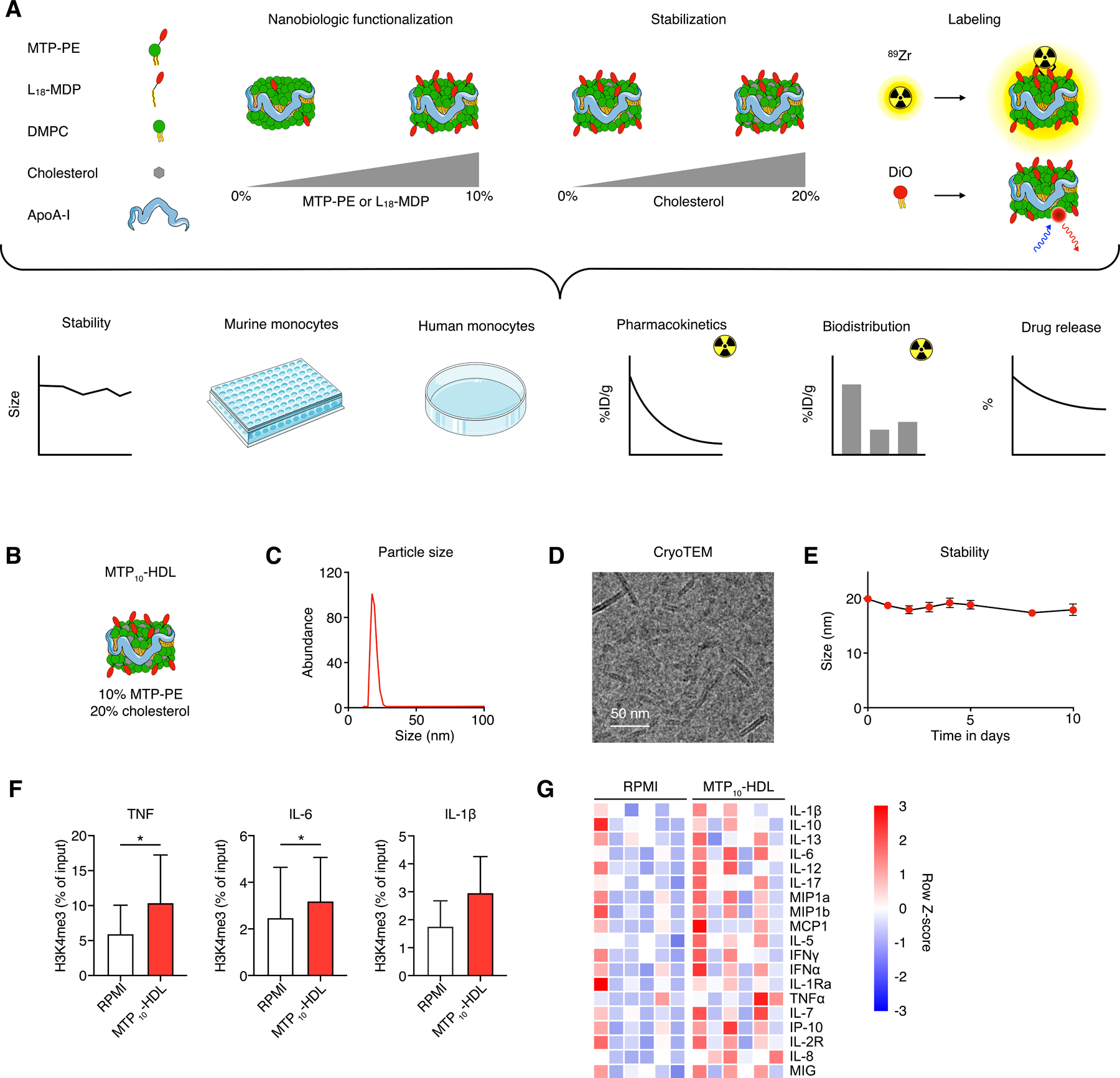
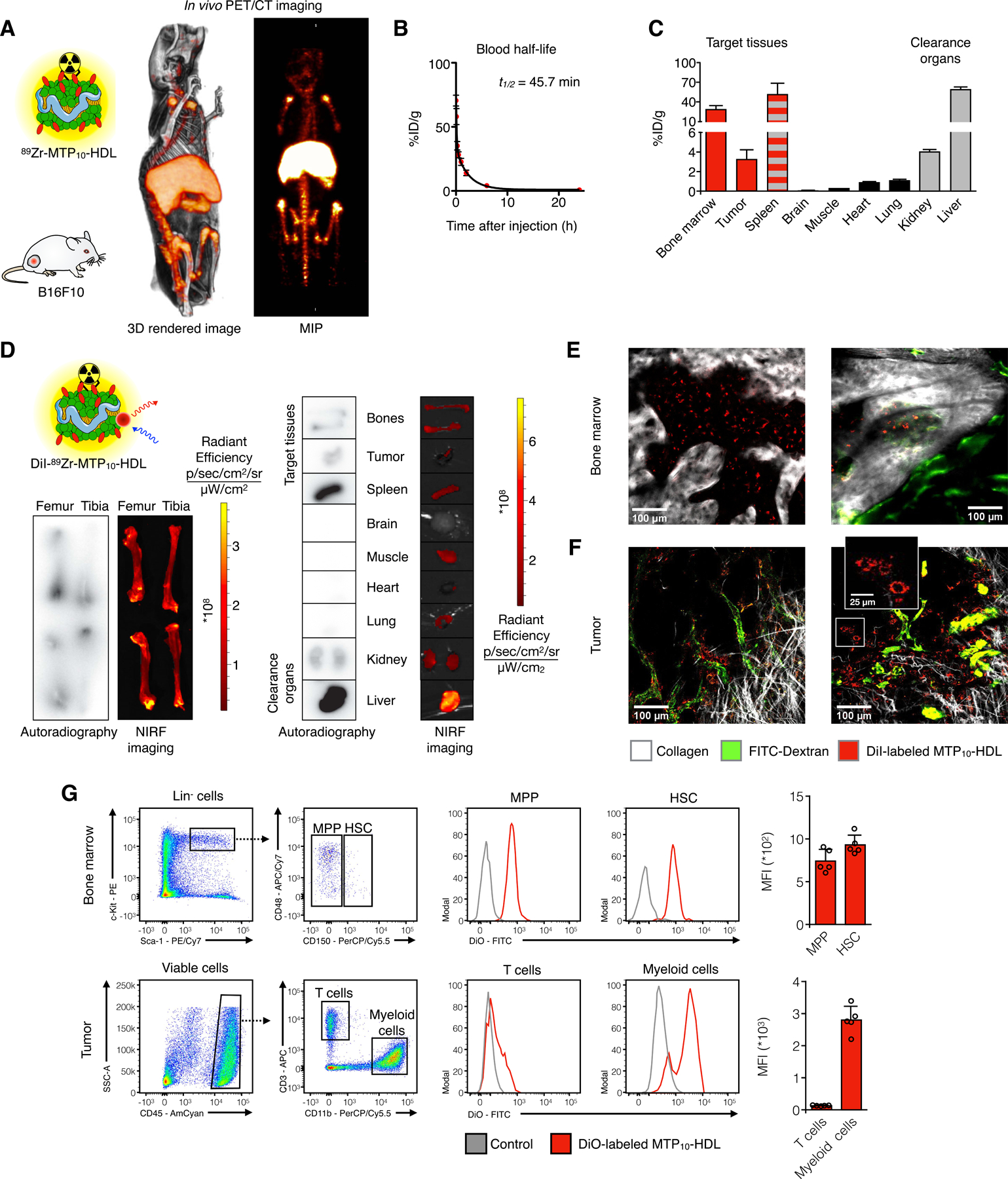
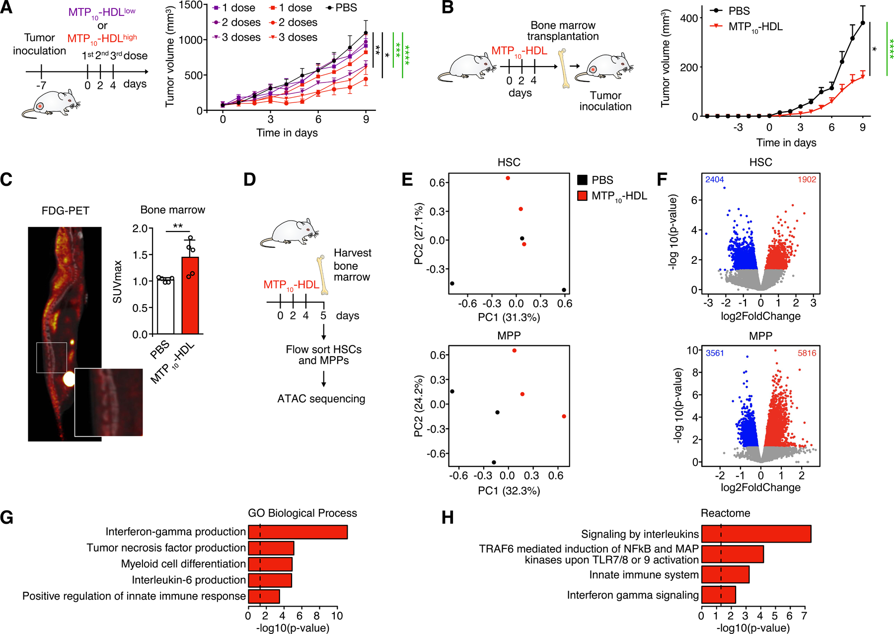
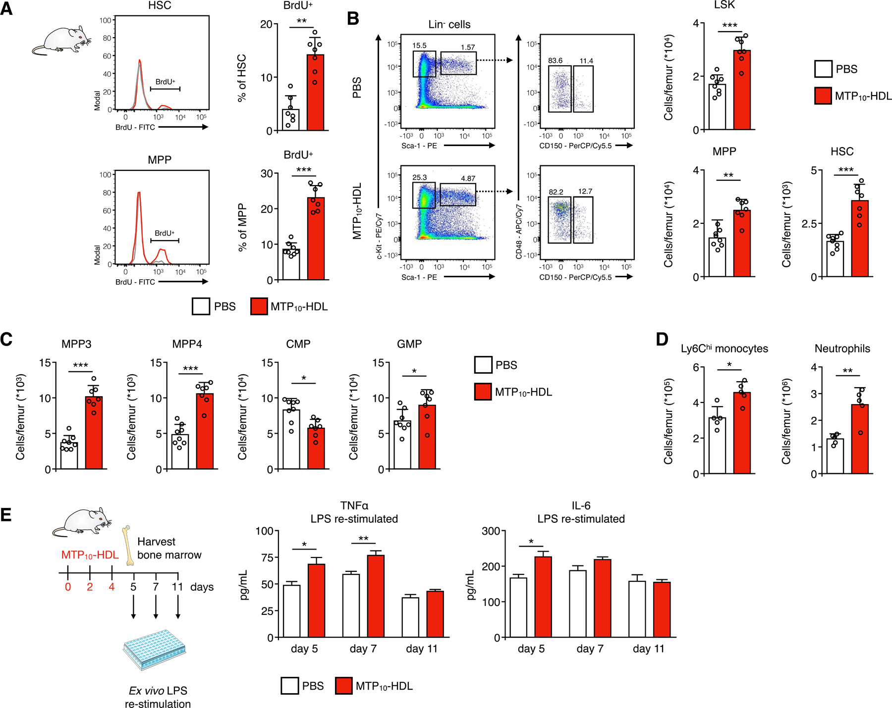
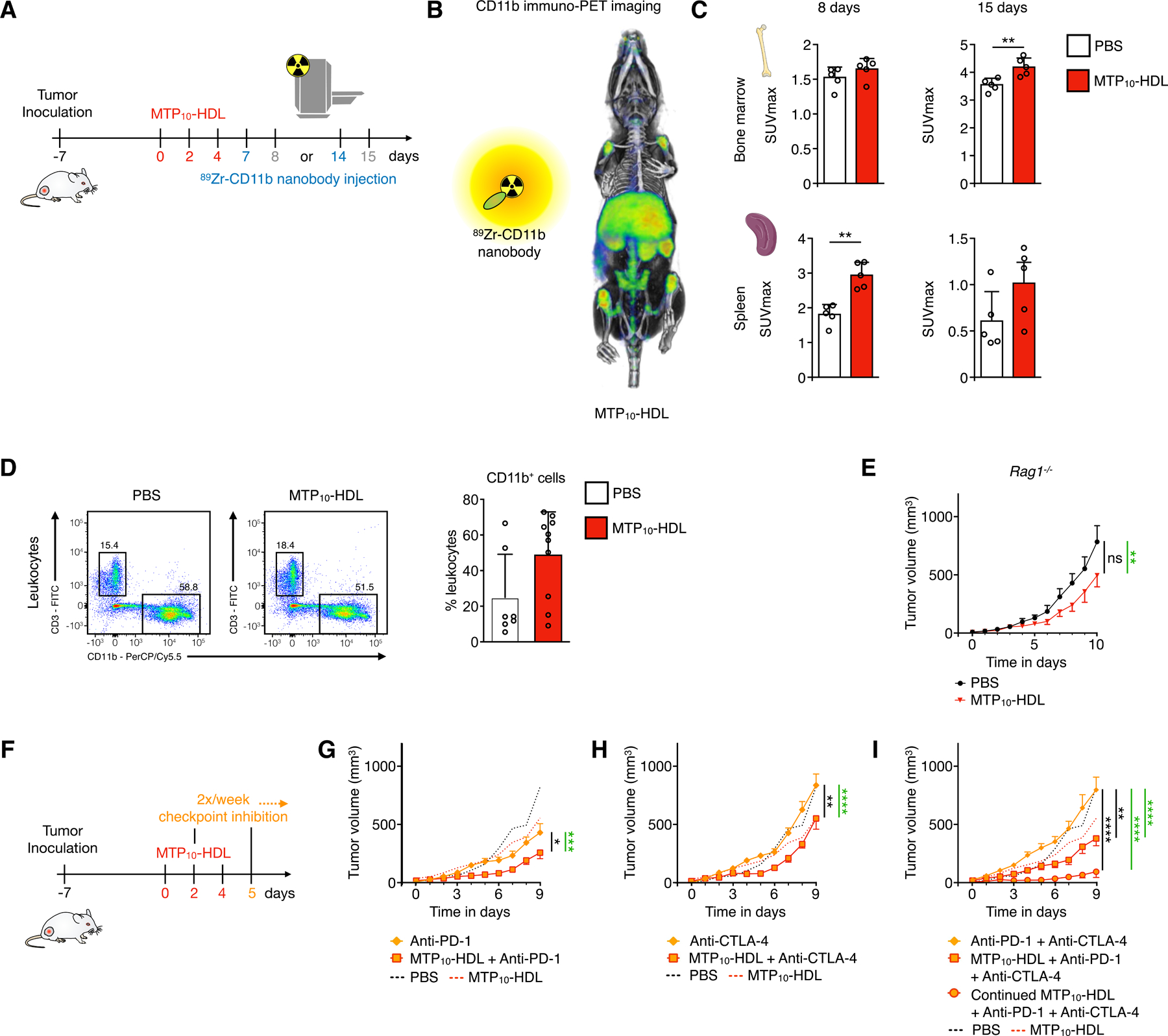
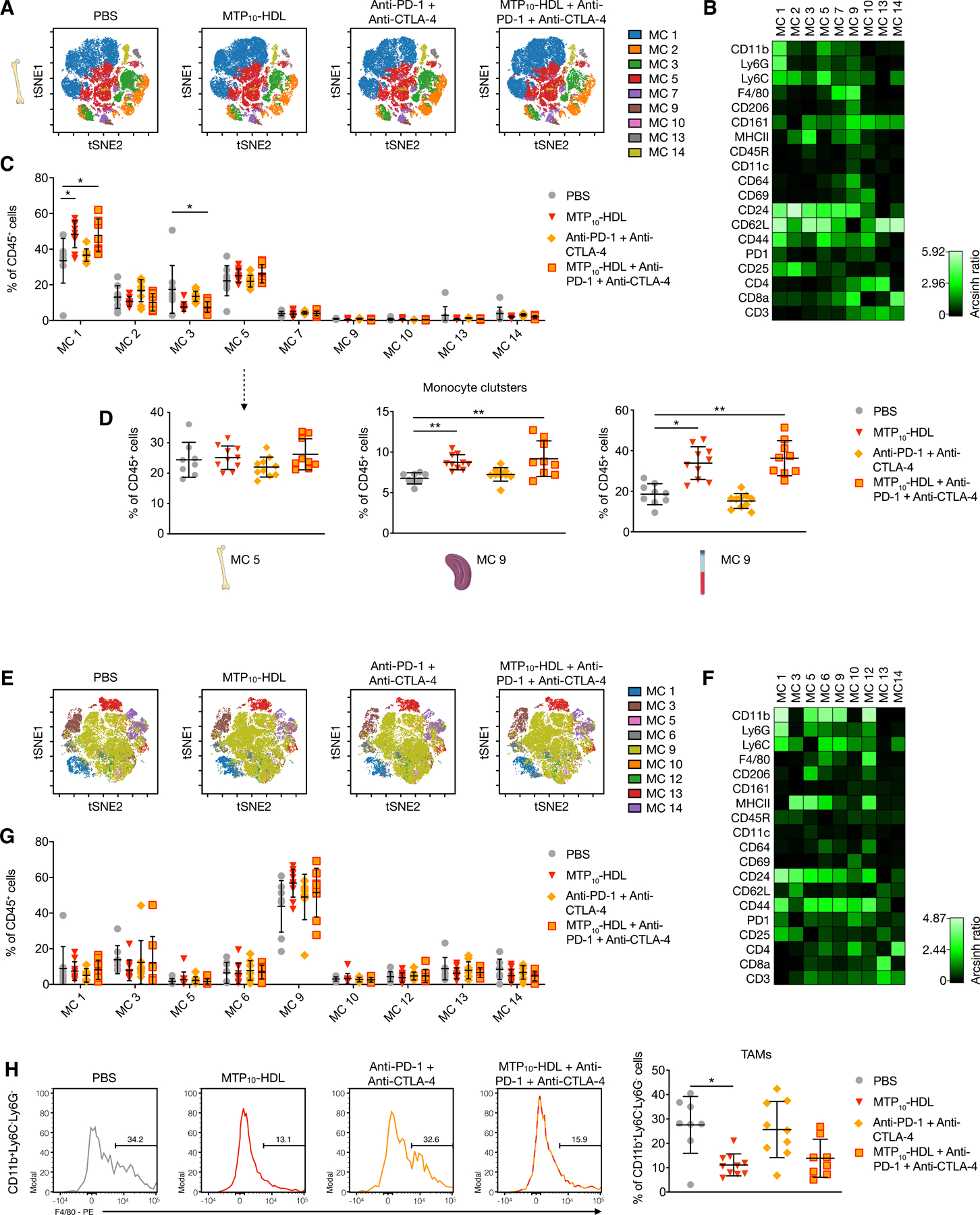
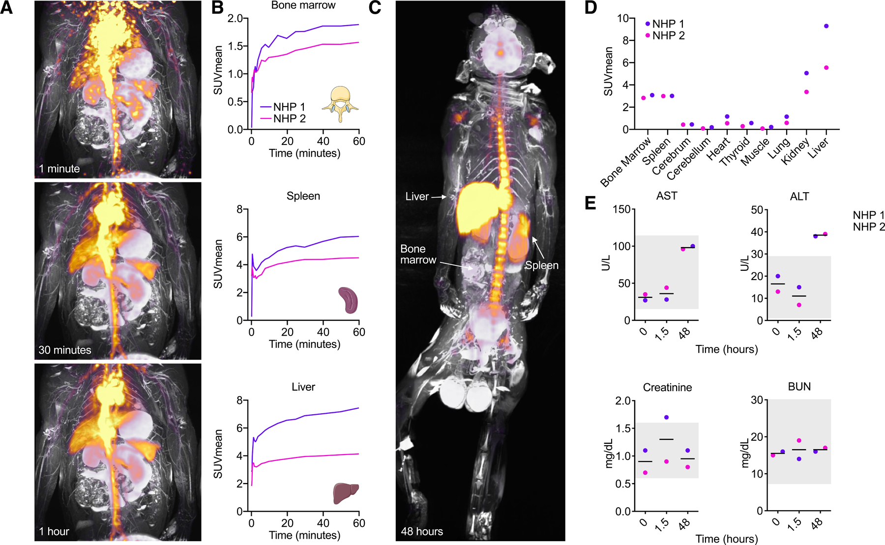
Trained immunity, a functional state of myeloid cells, has been proposed as a compelling immune-oncological target. Its efficient induction requires direct engagement of myeloid progenitors in the bone marrow. For this purpose, we developed a bone marrow-avid nanobiologic platform designed specifically to induce trained immunity. We established the potent anti-tumor capabilities of our lead candidate MTP10-HDL in a B16F10 mouse melanoma model. These anti-tumor effects result from trained immunity-induced myelopoiesis caused by epigenetic rewiring of multipotent progenitors in the bone marrow, which overcomes the immunosuppressive tumor microenvironment. Furthermore, MTP10-HDL nanotherapy potentiates checkpoint inhibition in this melanoma model refractory to anti-PD-1 and anti-CTLA-4 therapy. Finally, we determined MTP10-HDL's favorable biodistribution and safety profile in non-human primates. In conclusion, we show that rationally designed nanobiologics can promote trained immunity and elicit a durable anti-tumor response either as a monotherapy or in combination with checkpoint inhibitor drugs.

Keywords: cancer; checkpoint inhibitors; immunotherapy; innate immunity; melanoma; myeloid cells; nanobiologics; nanomedicine; nanotechnology; trained immunity.


Conflict of interest statement

Declaration of Interests W.J.M.M., L.A.B.J., J.O., Z.A.F., and M.G.N. are scientific co-founders of and have equity in Trained Therapeutix Discovery. W.J.M.M. and Z.A.F. have consulting agreements with Trained Therapeutix Discovery.

Figures

Figure 1.
Figure 1.. Nanobiologic screening and lead candidate selection
(A) (top) Schematic overview of nanobiologic library details. Individual nanobiologics are composed of human apoA1, the phospholipid DMPC and stabilized by cholesterol. Different surface densities of MDP or MTP are realized by varying the amount of L18-MDP or MTP-PE, respectively. The nanobiologics can be labeled with the radioisotope 89Zr or a fluorescent dye. (bottom) The different methods that were deployed to screen nanobiologics, integrating longitudinal size stability measurements by DLS, drug retention assays, murine and human monocyte in vitro training assays, pharmacokinetics and biodistribution studies in mice that received an intravenous injection of 89Zr-MTP10-HDL. (B) Schematic representation of the lead nanobiologic MTP10-HDL, consisting of 10 mol% MTP-PE and 20 mol% cholesterol relative to DMPC. (C) Particle size of MTP10-HDL, as determined by DLS, is 20 nm. (D) CryoTEM image of MTP10-HDL reveals a discoidal structure approximately 15 nm in diameter, with a thickness of 5 nm. (E) DLS stability assay demonstrate that MTP10-HDL size remain stable for at least 10 days. (F) ChIP-qPCR of human monocytes treated with MTP10-HDL and RPMI show increased H3K4 methylation on the TNFA, IL6 and IL1B promoters after MTP10-HDL treatment. (n=3) (G) Heatmap of human monocyte cytokines production after in vitro training. A general increase of pro-inflammatory cytokines after MTP10-HDL training is observed. (n=6) Data are presented as mean ± SD. P values were calculated using a Wilcoxon matched-pairs signed rank test, *p < 0.05. See also Figure S1 and S2.
Figure 2.
Figure 2.. In vivo behavior of the MTP10-HDL nanobiologic
(A) Whole-body 3D-rendered and maximum intensity projection (MIP) of PET/CT at 24 hours after injection of 89Zr-MTP10-HDL displayed high uptake in the bone marrow (femur, tibia, and spine), liver, and spleen. (B) 89Zr-MTP10-HDL has a blood half-life of 45.7 minutes. (n=5) (C) Gamma counting of tissues from C57BL/6 mice 24 hours after 89Zr-MTP10-HDL injection. A favorable uptake in the spleen and the bone marrow was observed. (n=5) (D) Ex vivo NIRF imaging and autoradiography 24 hours after injection of dual labeled DiI-89Zr-MTP10-HDL show high uptake in the liver, spleen, and bone marrow. Bone marrow uptake is concentrated at the proximal and distal ends of the bone, where the red marrow is located. (n=5) (E-F) Intravital microscopy of live animals C57BL/6 eight hours post DiI-MTP10-HDL administration. FITC-dextran was injected intravenously to display the vasculature. (E) Intravital microscopy image of a live mouse calvarium eight hours post DiI-MTP10-HDL administration. Clear DiI-MTP10-HDL uptake can be seen throughout the bone marrow. FITC-dextran was injected intravenously to display the vasculature. clearly show noticeable cellular uptake within bone marrow. (F) Intravital microscopy image of a live mouse tumor eight hours post DiI-MTP10-HDL administration shows DiI-MTP10-HDL distribution around the tumor vasculature. The inset shows uptake of DiI-MTP10-HDL in TAMs. FITC-dextran was injected intravenously to display the vasculature. (G) Flow cytometry of bone marrow and tumors 24 hours after DiO-MTP10-HDL administration. Identification of HSC and MPP (top) and T cells and myeloid cells (bottom) with representative histograms. Uptake was observed in bone marrow HSCs and MPPs as well as myeloid cells within the tumor, but not in T cells. (n=5) For all panels, data are presented as mean ± SD. See also Figure S3.
Figure 3.
Figure 3.. MTP10-HDL treatment inhibits tumor growth and activates HSCs
(A) In vivo tumor growth profiling in C57BL/6 mice inoculated with 1×10⁵ B16F10 tumor cells. Tumor growth curves of the different treatment groups are shown. Mice received either PBS or one, two, or three intravenous injections at either a low (0.375 mg/kg) or high (1.5 mg/kg) MTP10-HDL dose. A clear dose response was observed. (n=8–10 per group) Significance was calculated for tumor growth rate (black) and tumor size (green). (B) Tumor growth curves in bone marrow transplantation study. Naïve radiated mice received bone marrow from donors treated PBS or MTP10-HDL. Tumor inoculation of 1×10⁵ B16F10 cells was performed after a 6-week recovery period. A significant reduction in tumor growth was observed in mice that received bone marrow from mice treated with MTP10-HDL. (n=8–10 per group) Significance was calculated for tumor growth rate (black) and tumor size (green). (C) 18F-FDG-PET of C57BL/6 mice treated with MTP10-HDL. 18F-FDG was intravenously injected one hour before PET/CT imaging. A higher SUVmax of the bone marrow was observed in mice injected with MTP10-HDL, indicating increased metabolic activity. (n=5 per group) (D) Schematic overview of the performed ATAC sequencing experiments. C57BL/6 mice were treated with either PBS or MTP10-HDL. At day 5, bone marrow was harvested and sorted for HSCs and MPPs and these cells were subsequently subjected to ATAC sequencing. (E) Principle component analysis of ATAC-sequencing data shows clustering of different treatment conditions in MPPs and HSCs. (F) Volcano plot displaying open chromatin loci as determined by ATAC sequencing in HSCs (top) and MPPs (bottom). Average signal is represented as log2 fold change. Significantly up- (non-adj. p value < 0.05, log2FC > 1) and downregulated (non-adj. p value < 0.05, log2FC < −1) peaks are shown. (G-H) Overrepresented trained immunity-associated pathways that were upregulated in MPPs after MTP10-HDL treatment. Results from the Gene Ontology Biological Processes (G) and Reactome library (H) are displayed. Data are presented as mean ± SD and mean ± SEM for tumor growth experiments. P values were calculated using a Mann–Whitney U tests (two-sided) or an unpaired t-test (two-tailed). *p < 0.05, **p < 0.01, ***p < 0.001, ****p < 0.0001, ns= not significant. See also Figure S4.
Figure 4.
Figure 4.. MTP10-HDL treatment induces trained immunity in the bone marrow
Representative flow cytometry plots of bone marrow harvested from C57BL/6 mice treated with MTP10-HDL or PBS (A) BrdU proliferation assay. Mice treated with either MTP10-HDL or PBS received a BrdU injection 48 hours before euthanization, after which bone marrow was harvested. BrdU-positive hematopoietic stem cells (HSCs) and multipotent progenitors (MPPs) increased by 259% and 168%, respectively, indicating increased proliferation. (n=7–8 per group) (B-C) Representative flow cytometry plots of bone marrow harvested from C57BL/6 mice treated with MTP10-HDL or PBS and graphs showing the frequency of HSCs, MPPs, Lineage Sca1+ c-kit (LSK), MPP3, MPP4, granulocyte-monocyte progenitors (GMPs) significantly increases after MTP10-HDL. Whereas the amount of common myeloid progenitors (CMPs) significantly decreases. (n=7–8 per group) (D) Frequency of Ly6Chi monocyte and neutrophil counts significantly increases after MTP10-HDL treatment. (n=5 per group) (E) Cytokine concentrations in medium after restimulation of bone marrow cells harvested from C57BL/6 mice at day 5, 7 or 11 that were treated with either MTP10-HDL or PBS. At day 5, a significant increase in TNF-α and IL-6 was observed. (n=5–8 per group) Data are presented as mean ± SD. P values were calculated using a Mann–Whitney U tests (two-sided) or an unpaired t-test (two-tailed). *p < 0.05, **p < 0.01, ***p < 0.001, ****p < 0.0001, ns= not significant. See also Figure S5.
Figure 5.
Figure 5.. Inducing trained immunity potentiates checkpoint blockade therapy
(A-C) C57BL/6 mice inoculated with 1×10⁵ B16F10 cells treated with either MTP10-HDL or PBS received an intravenous injection of 89Zr-CD11b-NB at day seven or day 14. 89Zr-CD11b-NB was allowed to circulate for 24 hours before PET/CT imaging was performed. There is a higher SUVmax in the bone marrow and the spleen indicating higher amounts of CD11b-expressing cells present. (n=5 per group) (D) Flow cytometry gating strategy for tumors 24 hours after MTP10-HDL administration. Immune cells were isolated using Percoll gradient. A significant decrease in the amount of monocytes as a percentage of CD11b positive cells and an increase in neutrophils was observed. (n=7–10) (E) Rag1−/− mice inoculated with 1×10⁵ B16F10 cells were treated with either MTP10-HDL or PBS and tumor size was measured daily. Treatment with MTP10-HDL shows significant tumor inhibition but no inhibition of tumor growth rate. Significance was calculated for tumor growth rate (black) and tumor size (green). (n=10 per group) (F) Schematic overview of checkpoint inhibitor experiment. C57BL/6 mice inoculated with 1×10⁵ B16F10 cells were randomized into one of 7 treatment groups. PBS and MTP10-HDL results are shown in graphs’ dotted lines. Primary outcome was the comparison between checkpoint inhibitor immunotherapy alone versus in combination with MTP10-HDL. Significance was calculated for tumor growth rate (black) and tumor size (green). (G) Anti-PD-1 shows no significant tumor growth rate inhibition but does significantly decrease in tumor size. Adding MTP10-HDL significantly inhibits tumor growth rate as compared to anti-PD-1. (H) Anti-CTLA-4 does not significantly inhibit tumor growth rate or size, but combination with MTP10-HDL does significantly inhibit tumor growth rate and size. (I) Combining anti-PD-1 + anti-CTLA-4 has no significant effect on tumor growth rate and tumor size. Adding MTP10-HDL dramatically decreases the tumor growth rate, an effect that is even more pronounced after the MTP10-HDL regimen rises from three to six injections. Data are presented as mean ± SD and mean ± SEM for tumor growth experiments. P values were calculated using a Mann–Whitney U tests (two-sided) or an unpaired t-test (two-tailed). *p < 0.05, **p < 0.01, ***p < 0.001, ****p < 0.0001, ns= not significant.
Figure 6.
Figure 6.. Trained immunity causes a systemic shift towards myeloid cells
Tumor-bearing C57BL/6 mice were treated with PBS, MTP10-HDL, anti-CTLA-4 + anti-PD-1, or MTP10-HDL combined with anti-CTLA-4 + anti-PD-1. Leukocyte populations in the bone marrow, spleen, and blood were analyzed at day five. (A) Top panels show viSNE-plots from concatenated bone marrow samples per treatment group. (n=8–10 per group) Pie charts show cell distribution within each metacluster. Metaclusters containing less than 0.5% of total cells were excluded. (B) Heatmap shows the relative expression of different immune cell markers in each metacluster. Results were normalized by the row’s minimum. MC1: neutrophils, MC5: monocytes, MC13: CD4+ T cells, MC14: CD8+ T cells. (C) Quantification of cells within each metacluster as a percentage of CD45+ cells. Metacluster 1 shows a significant increase when MTP10-HDL was used as a treatment. (D) CD11b+, Ly6G, Ly6C+ metaclusters were selected from bone marrow, spleen, and blood. Marginal increase was observed in the bone marrow’s percentage of monocytes. However, spleen and blood show significant higher monocyte percentages than does control. (E) Top panels show viSNE-plots from all concatenated tumor samples. (F) Heatmap shows the relative expression of different immune cell markers in each metacluster. Results were normalized by the row’s minimum. (G) Percentage of CD45+ cells within each metacluster as a percentage of CD45+ cells. (H) Histograms of concatenated samples, per treatment group, showing F4/80 expression in CD11b+, Ly6C, Ly6G cells and quantification of F4/80+ TAMs per treatment group. Data are presented as mean ± SD. P values were calculated using a Mann–Whitney U tests (two-sided). *p < 0.05, **p < 0.01, ***p < 0.001, ****p < 0.0001, ns= not significant. See also Figure S6.
Figure 7.
Figure 7.. In vivo behavior of the MTP10-HDL nanobiologic in a non-human primate.
Two adult male non-human primates (Macaca fascicularis) were injected with 89Zr-MTP10-HDL at a dose of 0.0549 mg/kg and subjected to full body PET/MRI to investigate biodistribution. Blood measurements were done to investigate toxicity. (n=2) (A) Dynamic PET/MRI scans of a non-human primate 1, 30 and 60 minutes after injection of 89Zr-MTP10-HDL. Fast bone marrow and spleen accumulation, as well as liver uptake can be observed. (B) Organ SUVmean measurements over time revealed rapid uptake in the bone marrow, spleen and liver. (C) PET/MRI scan after 48 hours after 89Zr-MTP10-HDL injection displays a favorably high bone marrow and spleen accumulation relative to the liver. (D) Organ specific SUVmean after 48 hours shows high uptake in the bone marrow, spleen and liver. No uptake is found in the brain. (E) Blood chemistry performed on non-human primate serum taken at 0, 1.5 and 48 hours after 89Zr-MTP10-HDL administration. The grey box indicates reference values. ALT, AST, creatine, BUN levels show no signs of severe toxicity. Data are presented as mean ± SD. P values were calculated using a Mann–Whitney U tests (two-sided). *p < 0.05, **p < 0.01, ***p < 0.001, ****p < 0.0001, ns= not significant.

Comment in

Similar articles

Cited by

References

    1. Akira S, Uematsu S, and Takeuchi O (2006). Pathogen recognition and innate immunity. Cell 124, 783–801. - PubMed
    1. Amir el AD, Davis KL, Tadmor MD, Simonds EF, Levine JH, Bendall SC, Shenfeld DK, Krishnaswamy S, Nolan GP, and Pe’er D (2013). viSNE enables visualization of high dimensional single-cell data and reveals phenotypic heterogeneity of leukemia. Nat Biotechnol 31, 545–552. - PMC - PubMed
    1. Anderson KG, Stromnes IM, and Greenberg PD (2017). Obstacles Posed by the Tumor Microenvironment to T cell Activity: A Case for Synergistic Therapies. Cancer Cell 31, 311–325. - PMC - PubMed
    1. Arts RJ, Joosten LA, and Netea MG (2016). Immunometabolic circuits in trained immunity. Semin Immunol 28, 425–430. - PubMed
    1. Braza MS, van Leent MMT, Lameijer M, Sanchez-Gaytan BL, Arts RJW, Perez-Medina C, Conde P, Garcia MR, Gonzalez-Perez M, Brahmachary M, et al. (2018). Inhibiting Inflammation with Myeloid Cell-Specific Nanobiologics Promotes Organ Transplant Acceptance. Immunity 49, 819–828. - PMC - PubMed

Publication types

MeSH terms

LinkOut - more resources