. 2020 Oct 28;10(11):1974.
doi: 10.3390/ani10111974.

Swim Bladder Disorders in Koi Carp (Cyprinus carpio)

Affiliations

Swim Bladder Disorders in Koi Carp (Cyprinus carpio)

Rubina Sirri et al. Animals (Basel). .

Abstract

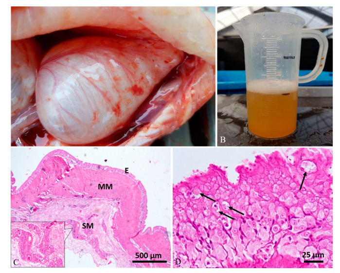
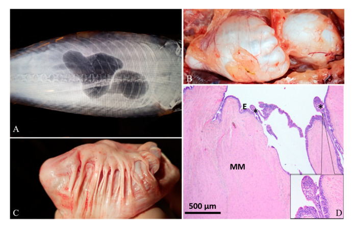
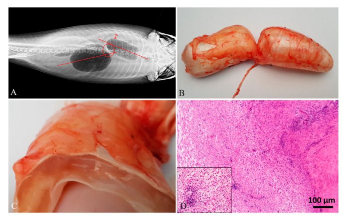
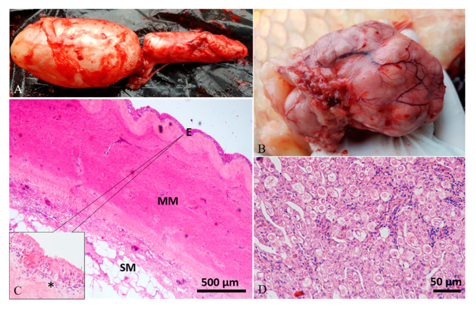
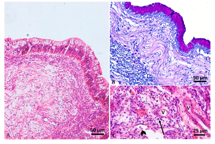
Swim bladder disorders and consequent buoyancy problems are encountered in ornamental fish, including koi carp. Nevertheless, beyond clinical and pharmacological management, they are largely underdiagnosed. In this study, nine koi carp showing abdominal swelling and abnormal swimming behavior were investigated. Clinical approach, varying from case to case, included ultrasonographic and X-ray investigations, bacteriological analysis of the collected fluid, antimicrobial susceptibility pattern, and possibly histological analysis. Diagnostic imaging, corroborating gross examination, documented swim bladder deformation/dislocation and serous fluid within the swim bladder chambers of most animals. Bacteria belonging to the Aeromonas hydrophila/caviae group and Shewanella xiamenensis were identified. S. xiamenensis strains showed a sensibility to all tested molecules except for one strain, which was resistant to tetracycline and cyprofloxacin. Antibiotic treatment succeeded in the full recovery of three cases in which S. xiamemensis infection was detected. Chronic aerocystitis was histologically documented where tissue was available. The swim bladder histopathological findings highlighted a chronic process that had compromised the quality of life of the animals. A multidisciplinary clinical-pathological and microbiological approach is highly suggested to recognize swim bladder conditions as early as possible, aiming to drive medical intervention and raising the chances of fish survival.

Keywords: cyprinid; diagnostic imaging; histopathology; koi carp; microbiology; ornamental fish; swim bladder disorders.


Conflict of interest statement

The authors declare no conflict of interest.

Figures

Figure 1
Figure 1
Case 4#. (A) Swim bladder showing a severe enlargement of chambers and multifocal superficial hyperemic streaks. (B) A conspicuous amount of yellow clear fluid was aspirated from the swim bladder. (C) Histological analysis showed severe thickening of the muscularis mucosae (MM) and perivascular inflammatory-cell infiltration in the submucosa (SM) (inset; hematoxylin–eosin (H&E) stain). (D) The mucosa (E) showed a severe epithelial hyperplasia with the presence of intracytoplasmic eosinophilic round aggregates compatible with parasitic stages (arrows) (H&E stain).
Figure 2
Figure 2
Case 5#. (A) Radiographic image of swim bladder showing abnormal shape. (B) Grossly, the chambers, especially the caudal one, showed an irregular surface. (C) At the opening, the internal surface was characterized by abundant fibrous connective tissue that twitched the swim bladder wall. (D) Histology shows a severe fibrous hyperplasia of the muscularis mucosae (MM) and a severe mucosal (E) hyperplasia forming papillae (asterisks), with abundant inflammatory cell exocytosis (inset; H&E stain).
Figure 3
Figure 3
Case 6#. (A) Radiographic image of swim bladder showing dislocation of the chambers. (B) Grossly, the size of the chambers appeared slightly enlarged. (C) When cut in a transversal section, abundant gelatinous material expanding the swim bladder layers was detected. (D) Histological analysis showed the muscularis mucosae expanded by abundant granulation tissue and a severe inflammatory cell infiltration, mainly composed of lymphocytes (inset; H&E stain).
Figure 4
Figure 4
Case 1#. (A) Swim bladder showing a moderate enlargement of caudal chamber. (B) The fish had a large intracoelomic tumor, which caused the compression of the swim bladder. (C) Histological analysis of the swim bladder showed fibrous hyperplasia of the muscularis mucosae (MM), submucosal (SM) multifocal inflammatory cell infiltration, edema of lamina propria (asterisk in inset), and mucosal (E) hyperplasia with squamous metaplasia (inset; H&E stain). (D) Histology of the tumor revealed a gonadal germ-cell tumor (H&E stain).
Figure 5
Figure 5
Case 2#. (A) Histology of swim bladder showing mucosal hyperplasia with mucous metaplasia and a severe mixed inflammatory cell infiltration expanding the lamina propria and muscularis mucosae (H&E stain). (B) Periodic acid–Schiff (PAS) stain highlights mucous metaplasia of epithelium. (C) The inflammatory reaction, composed of mainly lymphocytes (arrow) and mast cells (arrowhead), was located mainly around vessels (V) (H&E stain).

References

    1. Balon E.K. The oldest domesticated fishes, and the consequences of an epigenetic dichotomy in fish culture. J. Ichthyol. Aquat. Biol. 2006;11:47–86.
    1. Saint-Erne N. Diagnostic Techniques and Treatments for Internal Disorders of Koi (Cyprinus carpio) Veter. Clin. N. Am. Exot. Anim. Pr. 2010;13:333–347. doi: 10.1016/j.cvex.2010.05.012. - DOI - PubMed
    1. De Kock S., Gomelsky B. Biology and Ecology of Carp. Informa UK Limited; Boca Raton, FL, USA: 2015. Japanese Ornamental Koi Carp: Origin, Variation and Genetics; pp. 27–53.
    1. Sanders J. Aquatic Ambulatory Practice. Veter. Clin. N. Am. Exot. Anim. Pr. 2018;21:609–622. doi: 10.1016/j.cvex.2018.05.003. - DOI - PubMed
    1. Dey V.K. The global trade in ornamental fish. Info Fish Internat. 2016;4:52–55.