Review
. 2020 Oct 28;21(21):8019.
doi: 10.3390/ijms21218019.

Effect of Physico-Chemical Properties of Nanoparticles on Their Intracellular Uptake

Affiliations
Review

Effect of Physico-Chemical Properties of Nanoparticles on Their Intracellular Uptake

Parinaz Sabourian et al. Int J Mol Sci. .

Abstract

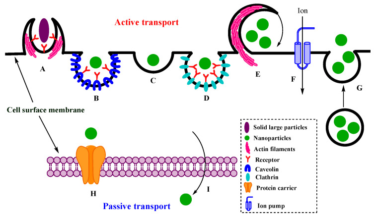
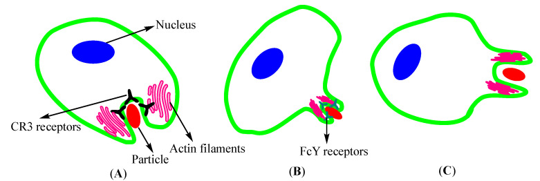
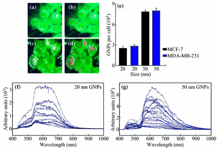
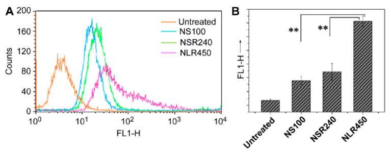
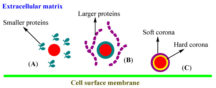
Cellular internalization of inorganic, lipidic and polymeric nanoparticles is of great significance in the quest to develop effective formulations for the treatment of high morbidity rate diseases. Understanding nanoparticle-cell interactions plays a key role in therapeutic interventions, and it continues to be a topic of great interest to both chemists and biologists. The mechanistic evaluation of cellular uptake is quite complex and is continuously being aided by the design of nanocarriers with desired physico-chemical properties. The progress in biomedicine, including enhancing the rate of uptake by the cells, is being made through the development of structure-property relationships in nanoparticles. We summarize here investigations related to transport pathways through active and passive mechanisms, and the role played by physico-chemical properties of nanoparticles, including size, geometry or shape, core-corona structure, surface chemistry, ligand binding and mechanical effects, in influencing intracellular delivery. It is becoming clear that designing nanoparticles with specific surface composition, and engineered physical and mechanical characteristics, can facilitate their internalization more efficiently into the targeted cells, as well as enhance the rate of cellular uptake.

Keywords: active/passive transport; cellular uptake; intracellular delivery; nanoparticles; physico-chemical properties.


Conflict of interest statement

The authors declare no conflict of interest.

Figures

Scheme 1
Scheme 1
Active and passive cell uptake of particles: (A) phagocytosis, (B) caveolin-mediated endocytosis, (C) clathrin–caveolin-independent endocytosis, (D) clathrin-mediated endocytosis, (E) macro-pinocytosis, (F) ion pumps, (G) exocytosis, (H) facilitated diffusion, and (I) simple diffusion.
Scheme 2
Scheme 2
Different mechanisms of nanoparticle (NP) phagocytosis: (A) CR3-mediated phagocytosis occurs by sinking NPs through CR3-receptors; (B) zipper mode phagocytosis via Fcγ-receptors including cell progression; (C) trigger mode phagocytosis with no receptors takes place through stimulating ruffles around particles.
Figure 1
Figure 1
Uptake of gold nanoparticles (GNPs). Hyperspectral image of cell uptake of (a) 20-nm- and (c) 50-nm-sized GNPs; (b,d) GNPs clusters mapped using reflectance spectra of GNPs; (e) GNP internalization per cell for 20- and 50-nm-sized GNPs in two different cell lines. (f,g) The reflectance spectra of the 20- and 50-nm-sized GNPs in the monolayers (a,c). Reprinted with permission from [55]. Copyright 2016 Springer Nature.
Figure 2
Figure 2
Quantification of the internalization of mesoporous silica NPs: fluorescence intensity (A), particle numbers (B), using fluorescent-activated cell sorting (FACS). ** Indicates statistical significance, p < 0.01). Reprinted with permission from [70]. Copyright 2010 Elsevier.
Figure 3
Figure 3
(A) Kinetics of cell uptake of fluorescently labeled silica NPs (25 μg/mL) in the complete (cMEM) and serum-free (SF) media, measured by flowcytometry; (B) curve in cMEM from A alone. Reprinted with permission from [77]. Copyright 2012 American Chemical Society.
Scheme 3
Scheme 3
Protein corona formation on the surface of NPs: (A) adsorption of smaller proteins to NP surface by rapid diffusion, (B) replacement of small proteins by larger ones, reconfiguration of proteins and final hard and soft corona formation (C).
Figure 4
Figure 4
Nanocapsular stiffness effects: (a) cellular uptake by RAW264.7 murine macrophages of surface-modified NPs; (b) fluorescence micrographs showing the cellular uptake of FA-PEG-modified NPs by CytD-treated SKOV3 cells. (** Indicates statistical significance, p < 0.01). Reprinted with permission from [114]. Copyright 2018 American Chemical Society.

References

    1. Jahromi L.P., Panah F.M., Azadi A., Ashrafi H. A mechanistic investigation on methotrexate-loaded chitosan-based hydrogel nanoparticles intended for CNS drug delivery: Trojan horse effect or not? Int. J. Biol. Macromol. 2019;125:785–790. doi: 10.1016/j.ijbiomac.2018.12.093. - DOI - PubMed
    1. Kumari N., Mathe V., Dongre P. Albumin nanoparticles conjugates binding with glycan-A strategic approach for targeted drug delivery. Int. J. Biol. Macromol. 2019;126:74–90. doi: 10.1016/j.ijbiomac.2018.12.184. - DOI - PubMed
    1. Soleimani M., Al Zaabi A.M., Merheb M., Matar R. Nanoparticles in Gene Therapy. Int. J. Integr. Biol. 2016;17:7–16.
    1. Kulkarni S.A., Feng S.-S. Effects of particle size and surface modification on cellular uptake and biodistribution of polymeric nanoparticles for drug delivery. Pharm. Res. 2013;30:2512–2522. doi: 10.1007/s11095-012-0958-3. - DOI - PubMed
    1. Ma N., Ma C., Li C., Wang T., Tang Y., Wang H., Mou X., Chen Z., He N. Influence of nanoparticle shape, size, and surface functionalization on cellular uptake. J. Nanosci. Nanotechnol. 2013;13:6485–6498. doi: 10.1166/jnn.2013.7525. - DOI - PubMed

LinkOut - more resources