Review
. 2020 Dec;43(12):965-979.
doi: 10.1016/j.tins.2020.10.002. Epub 2020 Oct 27.

Microglial Phagocytosis: A Disease-Associated Process Emerging from Alzheimer's Disease Genetics

Affiliations
Review

Microglial Phagocytosis: A Disease-Associated Process Emerging from Alzheimer's Disease Genetics

Anna Podleśny-Drabiniok et al. Trends Neurosci. 2020 Dec.

Abstract

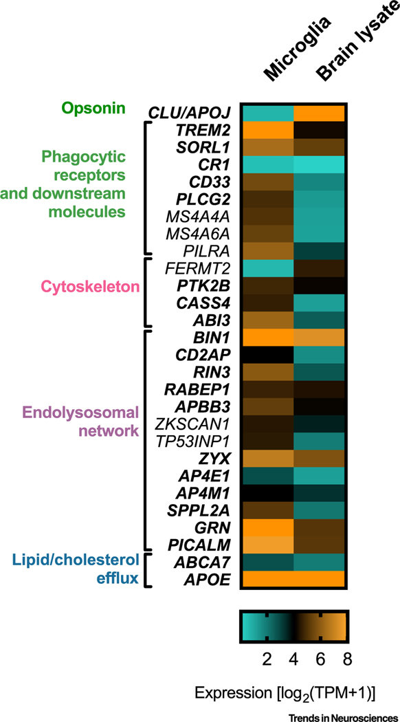
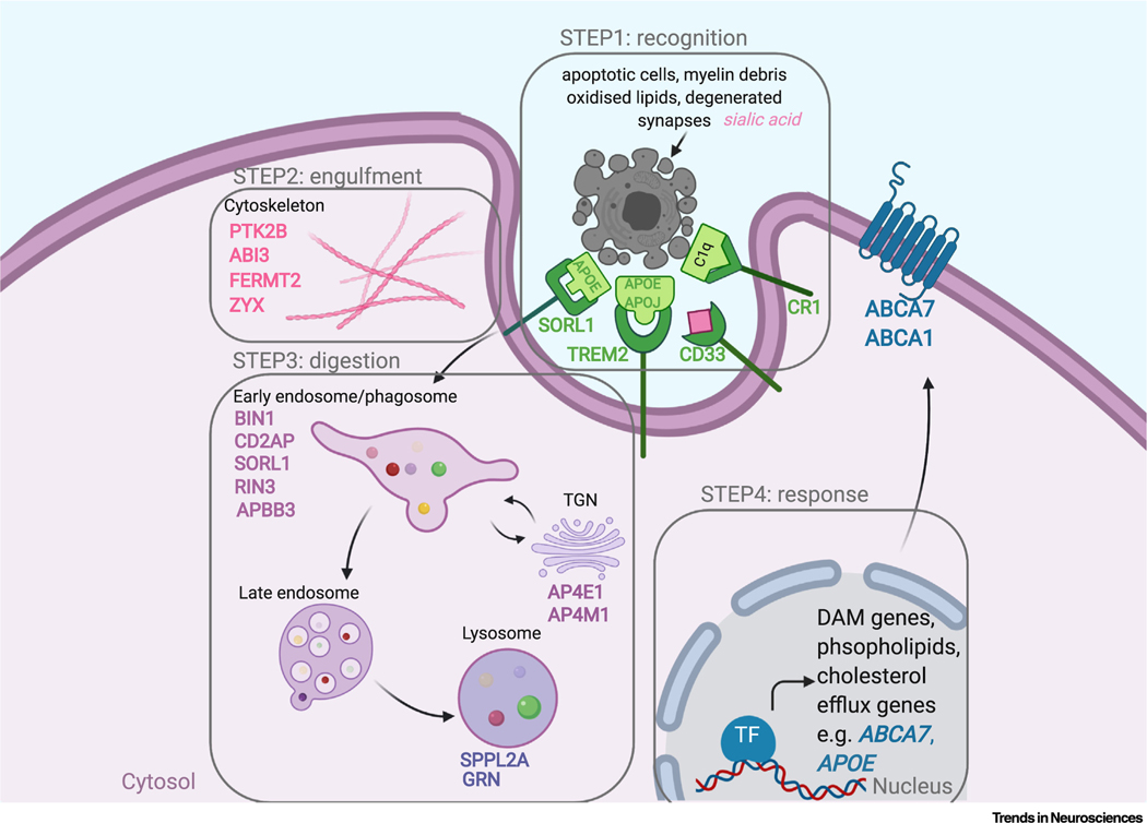
Alzheimer's disease (AD) is a debilitating, chronic neurodegenerative disease. Genetic studies involving genome-wide association studies (GWAS) and meta-analysis have discovered numerous genomic loci associated with AD; however, the causal genes and variants remain unidentified in most loci. Integration of GWAS signals with epigenomic annotations has demonstrated that AD risk variants are enriched in myeloid-specific enhancers, implicating myeloid cells in AD etiology. AD risk variants in these regulatory elements modify disease susceptibility by regulating the expression of genes that play crucial roles in microglial phagocytosis. Several of these AD risk genes are specifically expressed in myeloid cells, whereas others are ubiquitously expressed but are regulated by AD risk variants within myeloid enhancers in a cell type-specific manner. We discuss the impact of established AD risk variants on microglial phagocytosis and debris processing via the endolysosomal system.

Keywords: Alzheimer’s disease; GWAS; endolysosomal network; microglia; phagocytosis.


Conflict of interest statement

Disclaimer Statement A.M.G. has consulted for Eisai, Biogen, Pfizer, AbbVie, Cognition Therapeutics, and GSK; she also served on the Scientific Advisory Board of Denali Therapeutics (2015–2018).

Figures

Figure 1.
Figure 1.. Expression Pattern of Alzheimer’s Disease (AD) Risk Genes Involved in Phagocytosis.
Heatmap showing expression of selected AD risk genes in ex vivo human microglia and human brain lysates. Values of expression (transcripts per million, TPM) are plotted based on [28]. Genes marked in bold are discussed in this review.
Figure 2.
Figure 2.. Summary of Endolysosomal Processing in Microglial Cells, and Links to Candidate Alzheimer’s Disease (AD) Risk Genes.
Microglial cells constantly probe fragments of plasma membrane by endocytosis (top left) and large macromolecules (i.e., apoptotic bodies, myelin debris, synapses) by phagocytosis (top right). Inside the cell, engulfed material reaches early endosomes or phagosomes which mature and fuse with lysosomes (bottom, middle), forming a phagolysosome (bottom, right). Proteins are sorted to the trans-Golgi network (TGN) via retrograde transport and recycling endosomes. AD risk gene products are overlaid in cellular compartments they are known to regulate. Green indicates AD genes whose decreased expression is associated with increased AD risk; red indicates that increased expression is associated with increased AD risk. Gold indicates an unknown expression effect on AD risk. The expression directionality of AD risk genes is plotted based on [12] CD2AP, BIN1, RIN3, APPB3, ZYX, AP4E1, AP4M1, SPPL2A; [107] GRN; [98] RAB10; [62] CD33C; [64] CD33T; and [57] CR1 (CR1-S, CR1-F).
Figure 3.
Figure 3.. Candidate Alzheimer’s Disease (AD) Risk Genes in Microglial Phagocytosis.
Phagocytosis of apoptotic cells, myelin debris, synapses, and degenerated neurites is completed in four steps. Step 1: recognition [achieved by ‘find-me’ signals opsonized with apolipoprotein (APOE and APOJ, that are recognized by phagocytic receptors including TREM2 (triggering receptor expressed on myeloid cells 2), CR1 (complement receptor 1), and SORL1 (sortilin 1)]. Step 2: engulfment [achieved by cytoskeleton rearrangements regulated by e.g., PTK2B (protein tyrosine kinase 2β), ABI3]. Step 3: digestion [performed in the endolysosomal network and controlled by e.g., BIN1 (bridging integrator 1), CD2AP CD2-associated protein), RIN3 (Ras and Rab interactor 3), and SPPL2a (signal peptide peptidase-like 2a)]. Step 4: response [comprising activation of transcriptional program of clearance, i.e., DAM (disease/damage-associated microglia) genes and genes involved in phospholipids and cholesterol efflux, e.g., ABCA7, APOE]. AD risk genes are listed in each step and are overlaid in the cellular compartment whose function they control. Abbreviations: TF, transcription factor; TGN, trans-Golgi network.

References

    1. Alzheimer’s Association (2020) 2020 Alzheimer’s disease facts and figures. Alzheimers Dement. 16, 391–460 - PubMed
    1. Foley P (2010) Lipids in Alzheimer’s disease: a century-old story. Biochim. Biophys. Acta 1801, 750–753 - PubMed
    1. Holtzman DM et al. (2011) Alzheimer’s disease: the challenge of the second century. Sci. Transl. Med. 3, 77sr1 - PMC - PubMed
    1. Goate A et al. (1991) Segregation of a missense mutation in the amyloid precursor protein gene with familial Alzheimer’s disease. Nature 349, 704–706 - PubMed
    1. Clark RF et al. (1995) The structure of the presenilin 1 (S182) gene and identification of six novel mutations in early onset AD families. Nat. Genet. 11, 219–222 - PubMed