. 2021 Jan:102:155-162.
doi: 10.1016/j.ijid.2020.10.044. Epub 2020 Oct 28.

Cerebrospinal fluid findings in neurological diseases associated with COVID-19 and insights into mechanisms of disease development

Affiliations

Cerebrospinal fluid findings in neurological diseases associated with COVID-19 and insights into mechanisms of disease development

Otávio Melo Espíndola et al. Int J Infect Dis. 2021 Jan.

Abstract

Objectives: To analyze the cerebrospinal fluid (CSF) of patients with SARS-CoV-2 infection and neurological manifestations to provide evidence for the understanding of mechanisms associated with central nervous system (CNS) involvement in COVID-19.

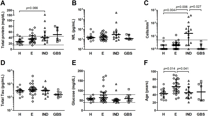
Methods: Patients (n = 58) were grouped according to their main neurological presentation: headache (n = 14); encephalopathy (n = 24); inflammatory neurological diseases, including meningoencephalitis (n = 4), acute myelitis (n = 3), meningitis (n = 2), acute disseminated encephalomyelitis (ADEM) (n = 2), encephalitis (n = 2), and neuromyelitis optica (n = 1); and Guillain-Barré syndrome (n = 6). Data regarding age, sex, cerebrovascular disease, and intracranial pressure were evaluated in combination with CSF profiles defined by cell counts, total protein and glucose levels, concentration of total Tau and neurofilament light chain (NfL) proteins, oligoclonal band patterns, and detection of SARS-CoV-2 RNA.

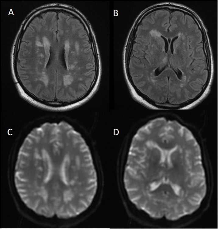
Results: CSF of patients with inflammatory neurological diseases was characterized by pleocytosis and elevated total protein and NfL levels. Patients with encephalopathy were mostly older men (mean age of 61.0 ± 17.6 years) with evidence of cerebrovascular disease. SARS-CoV-2 RNA in CSF was detected in 2 of 58 cases: a patient with refractory headache, and another patient who developed ADEM four days after onset of COVID-19 symptoms. Three patients presented intrathecal IgG synthesis, and four had identical oligoclonal bands in CSF and serum, indicating systemic inflammation.

Conclusion: Patients with neurological manifestations associated with COVID-19 had diverse CSF profiles, even within the same clinical condition. Our findings indicate a possible contribution of viral replication on triggering CNS infiltration by immune cells and the subsequent inflammation promoting neuronal injury.

Keywords: COVID-19; Cerebrospinal fluid; Neurofilament light protein; Oligoclonal bands; SARS-CoV-2; Total Tau protein.


Figures

Figure 1
Figure 1
CSF analysis of patients with COVID-19 and neurological manifestations. Patients were divided into four groups: headache (H); encephalopathy (E); inflammatory neurological diseases (IND), including acute disseminated encephalomyelitis, encephalitis, meningoencephalitis, meningitis, myelitis, and neuromyelitis optica; and Guillain-Barré syndrome (GBS). Comparison analysis was performed for (A) total protein, (B) neurofilament light chain (NfL), (C) cell counts, (D) total Tau protein, (E) glucose, and (F) age. Median values (line) and interquartile range (bars) are shown in (A, B, C, D, and E), and statistical analysis was performed with Dunn’s test with Bonferroni correction for multiple comparisons. Mean values (line) and standard deviation (bars) are shown in (F) and statistical analysis was performed with Bonferroni test for multiple comparisons. Differences with p-value < 0.05 were considered significant. Dashed lines indicate the reference values for (A) total protein (15–45 mg/dL), (C) CSF cell counts (<5 cells/mm3), and (E) glucose (40–80 mg/dL).
Figure 2
Figure 2
Correlation analysis of CSF data from patients with COVID-19. Correlation between CSF cell counts, total protein, glucose, Tau protein, and neurofilament light chain (NfL) levels was evaluated with Spearman’s rank of correlation in patients with SARS-CoV-2 infection and presenting with (A) headache (n = 14), (B) encephalopathy (n = 24), and (C) inflammatory neurological diseases (n = 14). Spearman’s rank correlation coefficient in bivariate analyses is represented by the color intensity of squares, which are shown in red for positive correlation and in blue for an inverse correlation. The size of squares corresponds to the level of statistical significance of the correlation (shown inside the squares), and associations with p-values < 0.05 were considered significant.
Figure 3
Figure 3
Characterization of neurological diseases associated with COVID-19. Qualitative parameters, such as age, sex, cerebrovascular disease (CVD), and elevated cerebrospinal fluid (CSF) total protein, and quantitative variables, including CSF cell counts, glucose, total Tau protein and neurofilament light chain (NfL) levels were evaluated by factor analysis of mixed data (FAMD) method. (A) Individuals (dots) were distributed into four main groups discriminated by colors according to their neurological outcome: headache, encephalopathy, inflammatory neurological diseases (IND), and Guillain-Barré syndrome (GBS). Correlation circles of quantitative variables (ellipses) and central points for the distribution of cases in a category (large dots) are shown for each group. The contribution of (B) qualitative and (C) quantitative variables to explain the neurological diseases associated with COVID-19 are shown.
Figure 4
Figure 4
Brain MRI of a patient with ADEM and detectable SARS-CoV-2 RNA in the cerebrospinal fluid. Hyperintense lesions on white matter substance in the deep hemispheric and periventricular areas both on (A, B) fluid-attenuated inversion recovery (FLAIR) and (C, D) apparent diffusion coefficient (ADC) map. Brain imaging was performed on hospital admission (day 1), four days after the onset of COVID-19 typical symptoms.

Similar articles

Cited by

References

    1. Abdi S., Ghorbani A., Fatehi F. The association of SARS-CoV-2 infection and acute disseminated encephalomyelitis without prominent clinical pulmonary symptoms. J Neurol Sci. 2020;416(June):117001. - PMC - PubMed
    1. Alberti P., Beretta S., Piatti M., Karantzoulis A., Piatti M.L., Santoro P. Guillain-Barré syndrome related to COVID-19 infection. Neurol Neuroimmunol Neuroinflamm. 2020;7(4) - PMC - PubMed
    1. Andersson M., Alvarez-Cermeño J., Bernardi G., Cogato I., Fredman P., Frederiksen J. Cerebrospinal fluid in the diagnosis of multiple sclerosis: a consensus report. J Neurol Neurosurg Psychiatry. 1994;57(August (8)):897–902. - PMC - PubMed
    1. Araujo A.Q.C., Silva M.T.T., Araujo A.P.Q.C. Zika virus-associated neurological disorders: a review. Brain. 2016;139(August (Pt 8)):2122–2130. - PubMed
    1. Arbour N., Day R., Newcombe J., Talbot P.J. Neuroinvasion by human respiratory coronaviruses. J Virol. 2000;74(October (19)):8913–8921. - PMC - PubMed