

A role for neuroimmune signaling in a rat model of Gulf War Illness-related pain
- PMID: 33127584
- PMCID: PMC7749855
- DOI: 10.1016/j.bbi.2020.10.022
A role for neuroimmune signaling in a rat model of Gulf War Illness-related pain
Abstract
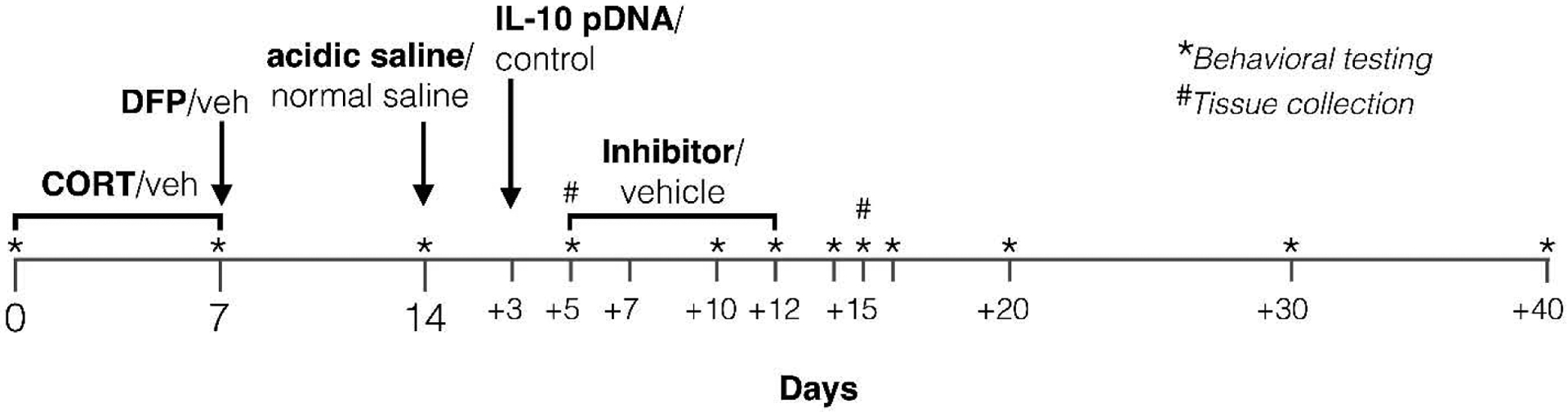
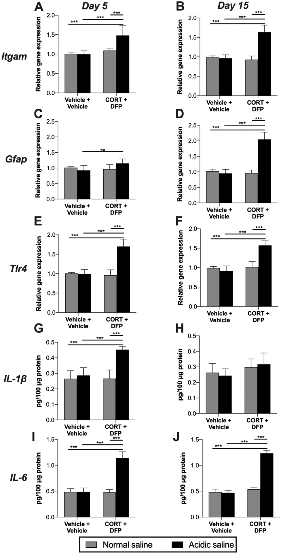
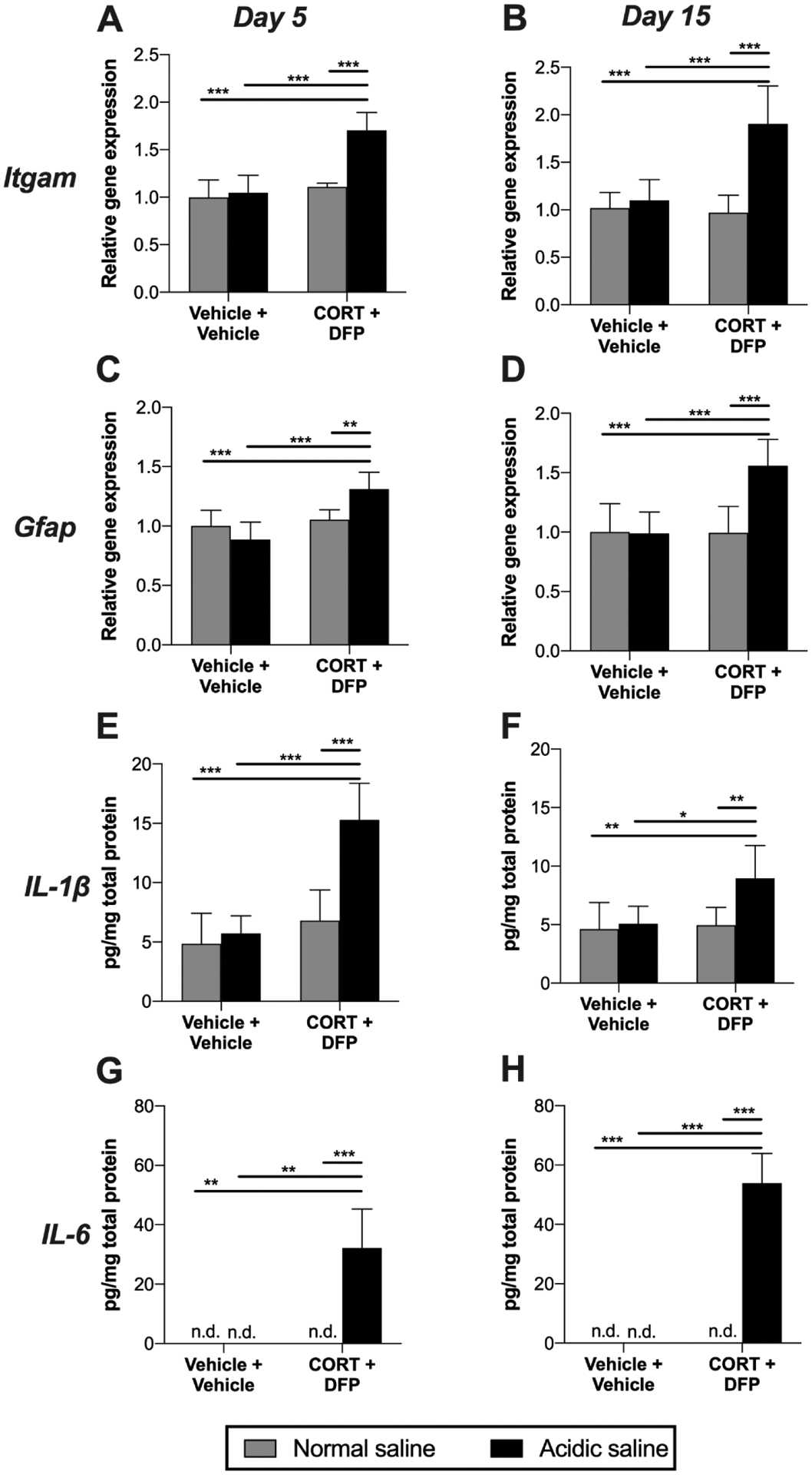
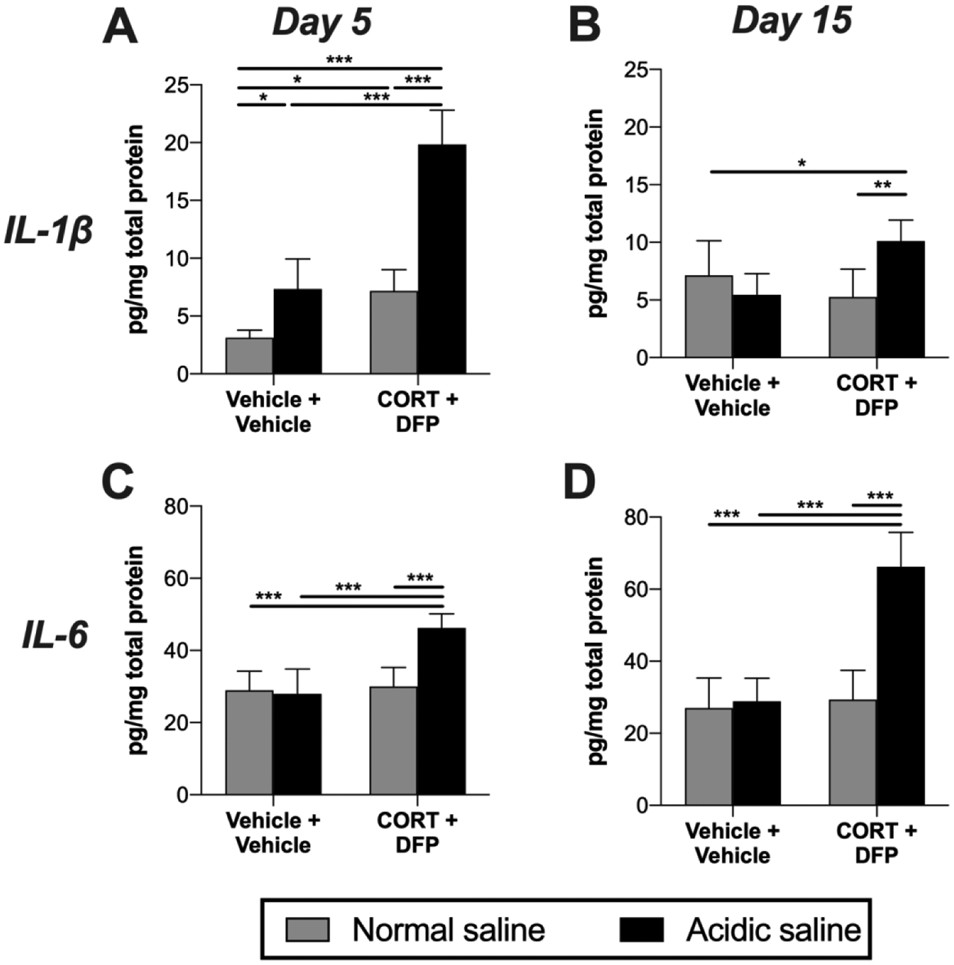
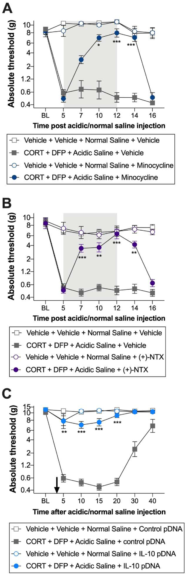
More than a quarter of veterans of the 1990-1991 Persian Gulf War suffer from Gulf War Illness (GWI), a chronic, multi-symptom illness that commonly includes musculoskeletal pain. Exposure to a range of toxic chemicals, including sarin nerve agent, are a suspected root cause of GWI. Moreover, such chemical exposures induce a neuroinflammatory response in rodents, which has been linked to several GWI symptoms in rodents and veterans with GWI. To date, a neuroinflammatory basis for pain associated with GWI has not been investigated. Here, we evaluated development of nociceptive hypersensitivity in a model of GWI. Male Sprague Dawley rats were treated with corticosterone in the drinking water for 7 days, to mimic high physiological stress, followed by a single injection of the sarin nerve agent surrogate, diisopropyl fluorophosphate. These exposures alone were insufficient to induce allodynia. However, an additional sub-threshold challenge (a single intramuscular injection of pH 4 saline) induced long-lasting, bilateral allodynia. Such allodynia was associated with elevation of markers for activated microglia/macrophages (CD11b) and astrocytes/satellite glia (GFAP) in the lumbar dorsal spinal cord and dorsal root ganglia (DRG). Additionally, Toll-like receptor 4 (TLR4) mRNA was elevated in the lumbar dorsal spinal cord, while IL-1β and IL-6 were elevated in the lumbar dorsal spinal cord, DRG, and gastrocnemius muscle. Demonstrating a casual role for such neuroinflammatory signaling, allodynia was reversed by treatment with either minocycline, the TLR4 inhibitor (+)-naltrexone, or IL-10 plasmid DNA. Together, these results point to a role for neuroinflammation in male rats in the model of musculoskeletal pain related to GWI. Therapies that alleviate persistent immune dysregulation may be a strategy to treat pain and other symptoms of GWI.
Keywords: Cytokines; Glia; Gulf War Illness; Immune cell; Neuroimmune; Pain; Sphingolipids.
Copyright © 2020 Elsevier Inc. All rights reserved.
Figures

Similar articles
-
Corticosterone potentiates DFP-induced neuroinflammation and affects high-order diffusion imaging in a rat model of Gulf War Illness.Brain Behav Immun. 2018 Jan;67:42-46. doi: 10.1016/j.bbi.2017.08.003. Epub 2017 Aug 4. Brain Behav Immun. 2018. PMID: 28782715 Free PMC article.
-
Corticosterone primes the neuroinflammatory response to DFP in mice: potential animal model of Gulf War Illness.J Neurochem. 2015 Jun;133(5):708-21. doi: 10.1111/jnc.13088. Epub 2015 Mar 24. J Neurochem. 2015. PMID: 25753028 Free PMC article.
-
Epigenetic impacts of stress priming of the neuroinflammatory response to sarin surrogate in mice: a model of Gulf War illness.J Neuroinflammation. 2018 Mar 17;15(1):86. doi: 10.1186/s12974-018-1113-9. J Neuroinflammation. 2018. PMID: 29549885 Free PMC article.
-
Acetylcholinesterase inhibitor exposures as an initiating factor in the development of Gulf War Illness, a chronic neuroimmune disorder in deployed veterans.Neuropharmacology. 2020 Jul;171:108073. doi: 10.1016/j.neuropharm.2020.108073. Epub 2020 Apr 2. Neuropharmacology. 2020. PMID: 32247728 Free PMC article. Review.
-
Pathophysiological basis and promise of experimental therapies for Gulf War Illness, a chronic neuropsychiatric syndrome in veterans.Psychopharmacology (Berl). 2023 Apr;240(4):673-697. doi: 10.1007/s00213-023-06319-5. Epub 2023 Feb 15. Psychopharmacology (Berl). 2023. PMID: 36790443 Review.
Cited by
-
Association of the tissue microstructural diffusivity and translocator protein PET in Gulf War Illness.Brain Behav Immun Health. 2021 Oct 6;18:100364. doi: 10.1016/j.bbih.2021.100364. eCollection 2021 Dec. Brain Behav Immun Health. 2021. PMID: 34693367 Free PMC article.
-
Dopamine and norepinephrine are embracing their immune side and so should we.Curr Opin Neurobiol. 2022 Dec;77:102626. doi: 10.1016/j.conb.2022.102626. Epub 2022 Sep 1. Curr Opin Neurobiol. 2022. PMID: 36058009 Free PMC article. Review.
-
Longitudinal Assessment of Ocular Biomarkers in Individuals With Gulf War Illness Symptoms.Mil Med. 2025 Jun 30;190(7-8):e1670-e1678. doi: 10.1093/milmed/usae457. Mil Med. 2025. PMID: 39361156 Free PMC article.
-
The β-adrenergic receptor blocker and anti-inflammatory drug propranolol mitigates brain cytokine expression in a long-term model of Gulf War Illness.Life Sci. 2021 Nov 15;285:119962. doi: 10.1016/j.lfs.2021.119962. Epub 2021 Sep 24. Life Sci. 2021. PMID: 34563566 Free PMC article.
-
FDA-approved cannabidiol [Epidiolex®] alleviates Gulf War Illness-linked cognitive and mood dysfunction, hyperalgesia, neuroinflammatory signaling, and declined neurogenesis.Mil Med Res. 2024 Aug 22;11(1):61. doi: 10.1186/s40779-024-00563-2. Mil Med Res. 2024. PMID: 39169440 Free PMC article.
References
-
- Abdullah L, Evans JE, Bishop A, Reed JM, Crynen G, Phillips J, Pelot R, Mullan MA, Ferro A, Mullan CM, Mullan MJ, Ait-Ghezala G, Crawford FC, 2012. Lipidomic Profiling of Phosphocholine Containing Brain Lipids in Mice with Sensorimotor Deficits and Anxiety-Like Features After Exposure to Gulf War Agents. Neuromol Med 14, 349–361. 10.1007/s12017-012-8192-z - DOI - PubMed
-
- Abdullah L, Evans JE, Montague H, Reed JM, Moser A, Crynen G, Gonzalez A, Zakirova Z, Ross I, Mullan C, Mullan M, Ait-Ghezala G, Crawford F, 2013. Chronic elevation of phosphocholine containing lipids in mice exposed to Gulf War agents pyridostigmine bromide and permethrin. Neurotoxicol Teratol 40, 74–84. 10.1016/j.ntt.2013.10.002 - DOI - PubMed
-
- Alhasson F, Das S, Seth R, Dattaroy D, Chandrashekaran V, Ryan CN, Chan LS, Testerman T, Burch J, Hofseth LJ, Horner R, Nagarkatti M, Nagarkatti P, Lasley SM, Chatterjee S, 2017. Altered gut microbiome in a mouse model of Gulf War Illness causes neuroinflammation and intestinal injury via leaky gut and TLR4 activation. PLoS ONE 12, e0172914 10.1371/journal.pone.0172914 - DOI - PMC - PubMed
-
- Ashbrook DG, Hing B, Michalovicz LT, Kelly KA, Miller JV, de Vega WC, Miller DB, Broderick G, O’Callaghan JP, McGowan PO, 2018. Epigenetic impacts of stress priming of the neuroinflammatory response to sarin surrogate in mice: a model of Gulf War illness. J Neuroinflammation 15, 86 10.1186/s12974-018-1113-9 - DOI - PMC - PubMed
Publication types
MeSH terms
Grants and funding
LinkOut - more resources
Full Text Sources
Other Literature Sources
Research Materials
Miscellaneous
