

Ischemic stroke disrupts the endothelial glycocalyx through activation of proHPSE via acrolein exposure
- PMID: 33127645
- PMCID: PMC7939480
- DOI: 10.1074/jbc.RA120.015105
Ischemic stroke disrupts the endothelial glycocalyx through activation of proHPSE via acrolein exposure
Abstract
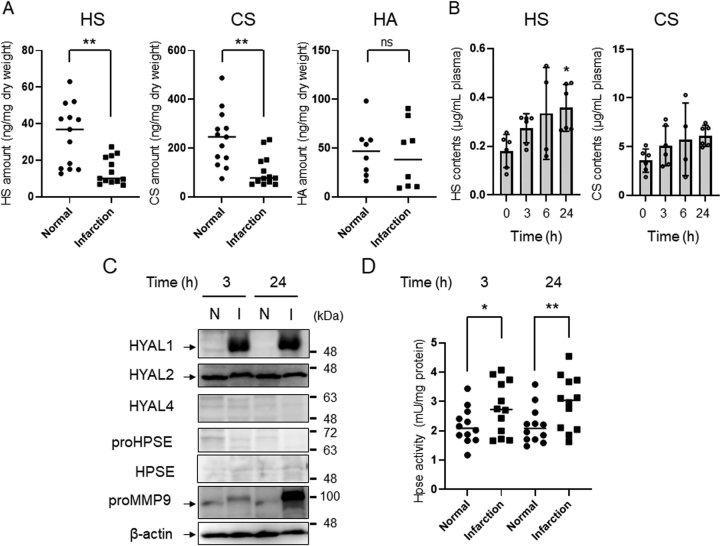
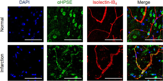
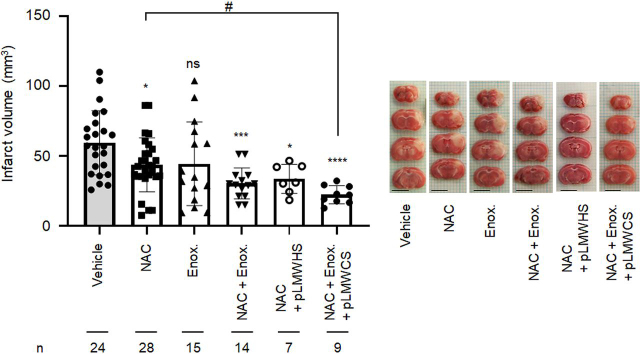
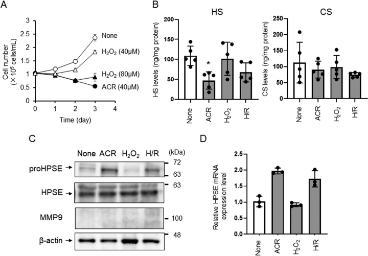
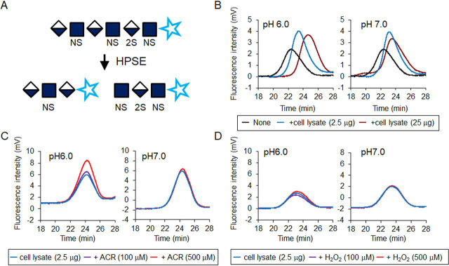
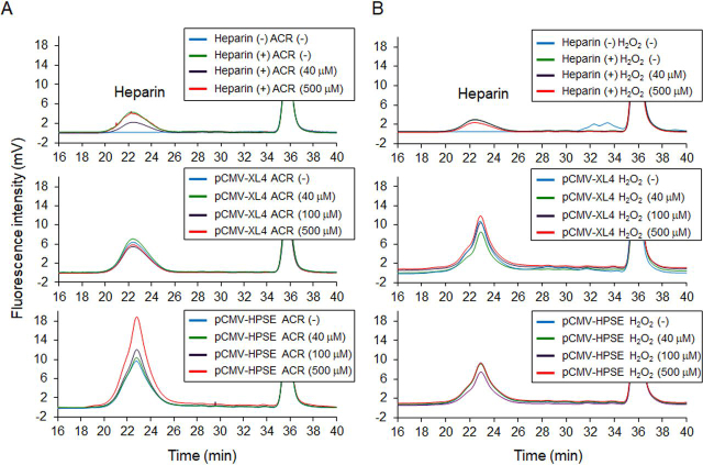
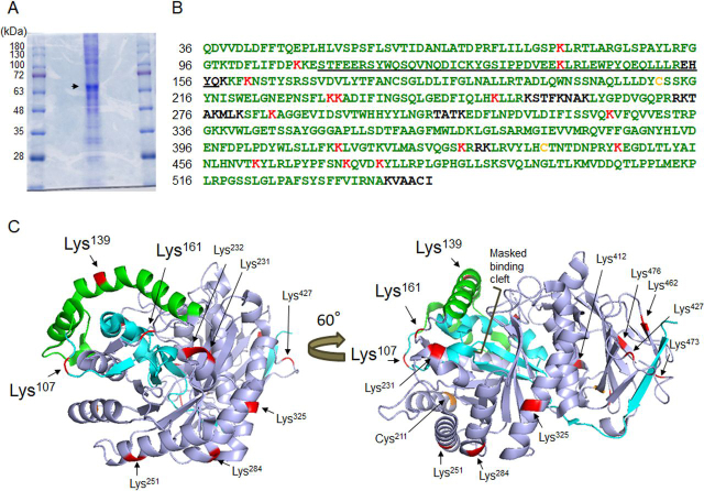
Infiltration of peripheral immune cells after blood-brain barrier dysfunction causes severe inflammation after a stroke. Although the endothelial glycocalyx, a network of membrane-bound glycoproteins and proteoglycans that covers the lumen of endothelial cells, functions as a barrier to circulating cells, the relationship between stroke severity and glycocalyx dysfunction remains unclear. In this study, glycosaminoglycans, a component of the endothelial glycocalyx, were studied in the context of ischemic stroke using a photochemically induced thrombosis mouse model. Decreased levels of heparan sulfate and chondroitin sulfate and increased activity of hyaluronidase 1 and heparanase (HPSE) were observed in ischemic brain tissues. HPSE expression in cerebral vessels increased after stroke onset and infarct volume greatly decreased after co-administration of N-acetylcysteine + glycosaminoglycan oligosaccharides as compared with N-acetylcysteine administration alone. These results suggest that the endothelial glycocalyx was injured after the onset of stroke. Interestingly, scission activity of proHPSE produced by immortalized endothelial cells and HEK293 cells transfected with hHPSE1 cDNA were activated by acrolein (ACR) exposure. We identified the ACR-modified amino acid residues of proHPSE using nano LC-MS/MS, suggesting that ACR modification of Lys139 (6-kDa linker), Lys107, and Lys161, located in the immediate vicinity of the 6-kDa linker, at least in part is attributed to the activation of proHPSE. Because proHPSE, but not HPSE, localizes outside cells by binding with heparan sulfate proteoglycans, ACR-modified proHPSE represents a promising target to protect the endothelial glycocalyx.
Keywords: acrolein; chondroitin sulfate; glycocalyx; heparan sulfate; heparanase; hyaluronan; hyaluronidase; ischemic stroke; stroke.
© 2020 Ko et al.
Conflict of interest statement
Conflict of interest—The authors declare that they have no conflicts of interest with the contents of this article.
Figures
Similar articles
-
Ulinastatin attenuates pulmonary endothelial glycocalyx damage and inhibits endothelial heparanase activity in LPS-induced ARDS.Biochem Biophys Res Commun. 2016 Sep 16;478(2):669-75. doi: 10.1016/j.bbrc.2016.08.005. Epub 2016 Aug 3. Biochem Biophys Res Commun. 2016. PMID: 27498004
-
Heparanase-induced endothelial glycocalyx degradation exacerbates lung ischemia/reperfusion injury in male mice.Physiol Rep. 2024 Oct;12(20):e70113. doi: 10.14814/phy2.70113. Physiol Rep. 2024. PMID: 39448392 Free PMC article.
-
Heparanase-Dependent Remodeling of Initial Lymphatic Glycocalyx Regulates Tissue-Fluid Drainage During Acute Inflammation in vivo.Front Immunol. 2019 Oct 4;10:2316. doi: 10.3389/fimmu.2019.02316. eCollection 2019. Front Immunol. 2019. PMID: 31636638 Free PMC article.
-
Endothelial Glycocalyx as a Shield Against Diabetic Vascular Complications: Involvement of Hyaluronan and Hyaluronidases.Arterioscler Thromb Vasc Biol. 2018 Jul;38(7):1427-1439. doi: 10.1161/ATVBAHA.118.310839. Epub 2018 Jun 7. Arterioscler Thromb Vasc Biol. 2018. PMID: 29880486 Free PMC article. Review.
-
Endothelial Glycocalyx Impairment in Disease: Focus on Hyaluronan Shedding.Am J Pathol. 2020 Apr;190(4):768-780. doi: 10.1016/j.ajpath.2019.11.016. Epub 2020 Feb 6. Am J Pathol. 2020. PMID: 32035885 Review.
Cited by
-
The Endothelial Glycocalyx: A Possible Therapeutic Target in Cardiovascular Disorders.Front Cardiovasc Med. 2022 May 13;9:897087. doi: 10.3389/fcvm.2022.897087. eCollection 2022. Front Cardiovasc Med. 2022. PMID: 35647072 Free PMC article. Review.
-
Surface charge, glycocalyx, and blood-brain barrier function.Tissue Barriers. 2021 Jul 3;9(3):1904773. doi: 10.1080/21688370.2021.1904773. Epub 2021 May 18. Tissue Barriers. 2021. PMID: 34003072 Free PMC article. Review.
-
The Neuroprotection of 1,2,4-Triazole Derivative by Inhibiting Inflammation and Protecting BBB Integrity in Acute Ischemic Stroke.CNS Neurosci Ther. 2024 Nov;30(11):e70113. doi: 10.1111/cns.70113. CNS Neurosci Ther. 2024. PMID: 39500736 Free PMC article.
-
Functional roles of polyamines and their metabolite acrolein in eukaryotic cells.Amino Acids. 2021 Oct;53(10):1473-1492. doi: 10.1007/s00726-021-03073-w. Epub 2021 Sep 21. Amino Acids. 2021. PMID: 34546444 Review.
-
Endothelial Dysfunction and Cardiovascular Disease: Hyperbaric Oxygen Therapy as an Emerging Therapeutic Modality?J Cardiovasc Dev Dis. 2024 Dec 19;11(12):408. doi: 10.3390/jcdd11120408. J Cardiovasc Dev Dis. 2024. PMID: 39728298 Free PMC article. Review.
References
-
- Zhang J., Takahashi H.K., Liu K., Wake H., Liu R., Maruo T., Date I., Yoshino T., Ohtsuka A., Mori S., Nishibori M. Anti-high mobility group box-1 monoclonal antibody protects the blood-brain barrier from ischemia-induced disruption in rats. Stroke. 2011;42:1420–1428. doi: 10.1161/STROKEAHA.110.598334. 21474801. - DOI - PubMed
-
- Shichita T., Hasegawa E., Kimura A., Morita R., Sakaguchi R., Takada I., Sekiya T., Ooboshi H., Kitazono T., Yanagawa T., Ishii T., Takahashi H., Mori S., Nishibori M., Kuroda K. Peroxiredoxin family proteins are key initiators of post-ischemic inflammation in the brain. Nat. Med. 2012;18:911–917. doi: 10.1038/nm.2749. 22610280. - DOI - PubMed
Publication types
MeSH terms
Substances
Associated data
- Actions
LinkOut - more resources
Full Text Sources
Medical
Molecular Biology Databases
