. 2020 Dec 25;295(52):18436-18448.
doi: 10.1074/jbc.RA120.015053. Epub 2020 Oct 30.

Development of a novel mammalian display system for selection of antibodies against membrane proteins

Affiliations

Development of a novel mammalian display system for selection of antibodies against membrane proteins

Nathan Robertson et al. J Biol Chem. .

Abstract

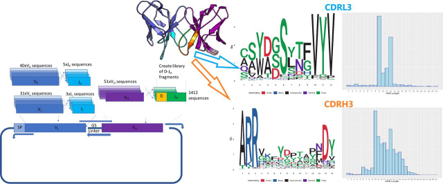
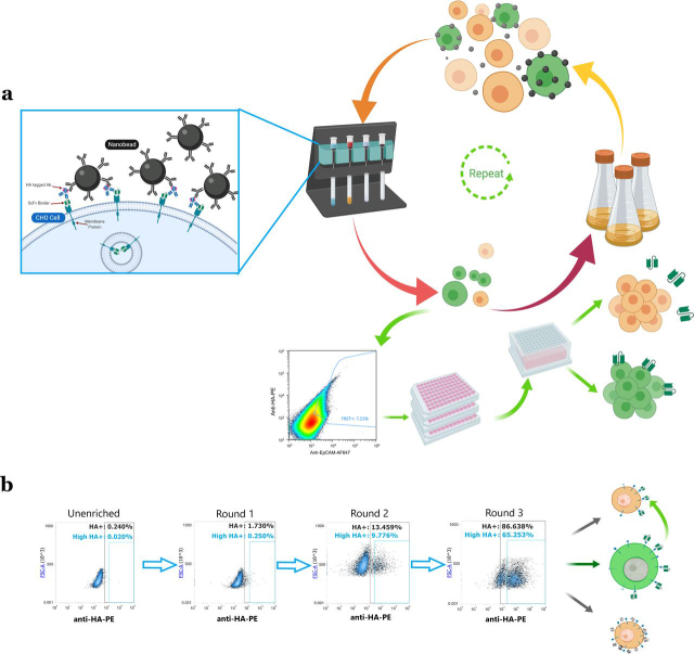
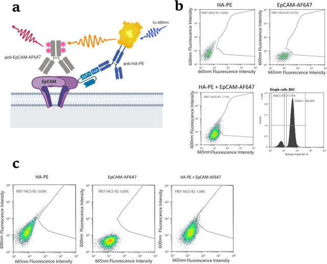
Reliable, specific polyclonal and monoclonal antibodies are important tools in research and medicine. However, the discovery of antibodies against their targets in their native forms is difficult. Here, we present a novel method for discovery of antibodies against membrane proteins in their native configuration in mammalian cells. The method involves the co-expression of an antibody library in a population of mammalian cells that express the target polypeptide within a natural membrane environment on the cell surface. Cells that secrete a single-chain fragment variable (scFv) that binds to the target membrane protein thereby become self-labeled, enabling enrichment and isolation by magnetic sorting and FRET-based flow sorting. Library sizes of up to 109 variants can be screened, thus allowing campaigns of naïve scFv libraries to be selected against membrane protein antigens in a Chinese hamster ovary cell system. We validate this method by screening a synthetic naïve human scFv library against Chinese hamster ovary cells expressing the oncogenic target epithelial cell adhesion molecule and identify a panel of three novel binders to this membrane protein, one with a dissociation constant (KD ) as low as 0.8 nm We further demonstrate that the identified antibodies have utility for killing epithelial cell adhesion molecule-positive cells when used as a targeting domain on chimeric antigen receptor T cells. Thus, we provide a new tool for identifying novel antibodies that act against membrane proteins, which could catalyze the discovery of new candidates for antibody-based therapies.

Keywords: antibody; antibody engineering; chimeric antigen receptor T cells (CAR-T); epithelial cell adhesion molecule (EpCAM); immunotherapy; mammalian display; membrane protein; therapeutic antibody discovery.


Conflict of interest statement

Conflict of interest—The authors are employees and shareholders of OXGENE, a biotech CRO company involved in the discovery and development of antibody-based therapeutics.

Figures

Figure 1
Figure 1
Flow cytometry analysis of CHO–EpCAM cells.a, cells stained with anti-EpCAM FITC in the absence or presence of doxycycline. b, cells transduced with lentiviruses expressing a control anti-EpCAM scFv with an HA tag stained with anti-HA-PE antibody in the absence or presence of doxycycline, i.e. demonstrating self-labeling.
Figure 2
Figure 2
Schematic overview of the construct of the scFv CHESS library, showing the numbers of V(D)J fragments recombined and shuffled into a lentiviral transfer plasmid. Natural germ-like variability was observed by NGS analysis of the CDR regions, the most variable being the CDR3 of the light and heavy chain. LOGO plots of the most common CDR3 length from these regions are shown alongside a histogram distribution of CDR3 lengths (right).
Figure 3
Figure 3
a, schematic of nanobead labeling, and overview of selection procedure. Images created with BioRender. b, flow cytometry scatter plots of cell outputs from three rounds of MACS selections showing anti-HA-PE staining. The enriched CHO cell culture contains three populations as shown in the schematic (right panel): cross-labeled cells, off-target self-labeled cells and target antigen-specific self-labeled cells.
Figure 4
Figure 4
FRET analysis of MACS-enriched cell pools after three rounds of selection.a, schematic of the FRET generation between the donor and acceptor fluorophores upon antigen binding by an scFv library member. b, scatter plots of the MACS-enriched CHO cells labeled with anti-HA-PE only (top left panel), anti-EpCAM-AF647 only (top right panel), and double labeling to generate FRET signal (bottom left panel). The histogram plot (bottom right panel) shows MACS-enriched cells separated from CMAC stained decoy cells (cells mixed at a ratio of 1:9). c, scatter plots showing the sorting gate used in the second round of FACS. The left and middle panels show the single stained controls, whereas the right panel shows the double stained (sorted) cell sample.
Figure 5
Figure 5
EpCAM-binding analysis of selected anti-EpCAM sequences that had been recloned and expressed either as scFv or as whole IgG1.a, flow cytometry analysis of binding of both scFv (left panel) and whole IgG (right panel) to CHO–EpCAM cells and CHO-X control cells. b, single-cycle kinetics SPR sensorgrams of antibodies formatted as whole IgG1 and captured on protein A sensor chips binding to EpCAM-ECD. The data from the curve fitting is shown in Table 1. SPR traces show representative data from four independent experiments.
Figure 6
Figure 6
Assessment of antigen specificity of CAR-T cells generated with the three selected antibodies.a, induction of activation marker CD25 on PBMC-derived CAR-T cells in co-culture with CHO–EpCAM or CHO-X cells (co-cultured at a ratio of 5:1) as measured by flow cytometry after 48 h of culture. b, cytotoxicity of PBMC-derived CAR-T cells when co-cultured with CHO-X or CHO–EpCAM as assessed by release of LDH after 48 h of culture. c and d, xCELLigence analysis of CHO-X (c) or CHO–EpCAM (d) cells co-cultured with PBMC-derived CAR-T cells (at a ratio of 1:5) monitored over 120 h. The data were measured in three biological triplicates where the mean is represented in solid line with the standard error shown by the dotted line of the same color. Statistical analysis was performed by two-way analysis of variance with Bonferroni post hoc tests and significance were assessed versus T cell cultured alone or CHO cells. ***, P < 0.001. e, assessment of Jurkat CAR-T cell activation by flow cytometry analysis of induction of activation marker CD69 after 4 h of co-culture with CHO–EpCAM, MCF7, or CHO-X cells (at a ratio of 1:1). The graph shows representative data from three independent experiments and shows the means and S.E. Statistical analysis was performed by two-way analysis of variance using Tukey's multiple comparison test for CHO–EpCAM or MCF7 cells compared with CHO-X cells. ****, P < 0.0001.

References

    1. Ho M., Nagata S., Pastan I. Isolation of anti-CD22 Fv with high affinity by Fv display on human cells. Proc. Natl. Acad. Sci. U.S.A. 2006;103:9637–9642. doi: 10.1073/pnas.0603653103. 16763048. - DOI - PMC - PubMed
    1. Akamatsu Y., Pakabunto K., Xu Z., Zhang Y., Tsurushita N. Whole IgG surface display on mammalian cells: application to isolation of neutralizing chicken monoclonal anti-IL-12 antibodies. J. Immunol. Methods. 2007;327:40–52. doi: 10.1016/j.jim.2007.07.007. 17719061. - DOI - PubMed
    1. Zhou C., Jacobsen F.W., Cai L., Chen Q., Shen W.D. Development of a novel mammalian cell surface antibody display platform. mAbs. 2010;2:508–518. doi: 10.4161/mabs.2.5.12970. 20716968. - DOI - PMC - PubMed
    1. Zhou Y., Chen Z.-R., Li C.-Z., He W., Liu S., Jiang S., Ma W.-L., Tan W., Zhou C. A novel strategy for rapid construction of libraries of full-length antibodies highly expressed on mammalian cell surfaces. Acta Biochim. Biophys. Sinica. 2010;42:575–584. doi: 10.1093/abbs/gmq055. - DOI - PubMed
    1. Parola C., Neumeier D., Friedensohn S., Csepregi L., Di Tacchio M., Mason D.M., Reddy S.T. Antibody discovery and engineering by enhanced CRISPR–Cas9 integration of variable gene cassette libraries in mammalian cells. mAbs. 2019;11:1367–1380. doi: 10.1080/19420862.2019.1662691. 31478465. - DOI - PMC - PubMed

Publication types