Case Reports
. 2020 Oct 30;13(10):e236926.
doi: 10.1136/bcr-2020-236926.

Mandibular odontogenic myxoma in a paediatric patient

Affiliations
Case Reports

Mandibular odontogenic myxoma in a paediatric patient

Raymond Patrick Shupak et al. BMJ Case Rep. .

Abstract

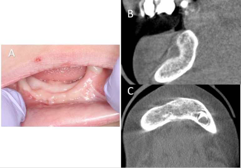
Odontogenic myxoma (OM) are benign, locally aggressive tumours that are rarely found in the paediatric maxillofacial region. OMs derive from mesenchymal odontogenic tissue. We describe the management of a 3-year-old girl who presented with a large right-sided mandibular lesion. Her treatment included conservative excision, curettage and peripheral ostectomy. A literature review was performed which calls into question the dogmatic practice of resection with 1 to 1.5 cm margins. Treatment approaches to the OM could potentially be altered in the paediatric patient.

Keywords: dentistry and oral medicine; ear; mouth; nose and throat/otolaryngology; oral and maxillofacial surgery; pathology.


Conflict of interest statement

Competing interests: None declared.

Figures

Figure 1
Figure 1
(A–C) Coronal, sagittal and axial slices of the preoperative maxillofacial CT scan showing a large, well-circumscribed radiolucent lesion that perforates the facial cortex (yellow arrow), is expansile, crosses midline and resorbs nearby dental root structures (white arrows). (D) 3D reconstruction.
Figure 2
Figure 2
Axial slice of a contrast-enhanced maxillofacial MRI demonstrating low T1 signal (A) to intermediate-to-bright T2 signal (B), with overall robust solid enhancement throughout suggesting this lesion is solid rather than cystic.
Figure 3
Figure 3
(A and B) H&E-stained lesion showing tumour specimen that is composed of bland, spindled-to-stellate uniform cells in a fibrous-to-myxoid background. There is no necrosis, mitoses or atypia suggestive of malignancy. (C) High power H&E staining of the specimen demonstrating haphazardly arranged spindle cell proliferation in an abundant mucoid-rich extracellular matrix interspersed with thin collagen fibres characteristic of odontogenic myxoma.
Figure 4
Figure 4
(A) Intraoperative intraoral view with nodular tissue expressing through the previous incisional biopsy site (yellow arrow). (B) View of the lesion after unroofing of the thin mandible facial cortex. (C and D) View of odontogenic myxoma specimen excised with associated extracted teeth.
Figure 5
Figure 5
(A) Mandible cavity after tumour excision and teeth extraction. (B) Peripheral ostectomy with round diamond bur (green arrow). (C) View of transected right inferior alveolar nerve in micro-forceps (yellow arrow). (D) Primary nerve repair (blue arrow).
Figure 6
Figure 6
(A) Preoperative photos demonstrating lower right facial asymmetry. (B) Postoperative images (6 weeks) with restoration of lower facial symmetry.
Figure 7
Figure 7
(A) Postoperative clinical images at 6 months. (B and C) Six-month postoperative CT images (sagittal and axial) showing bone regeneration without evidence of lesion recurrence.

References

    1. Thoma KH, Goldman HM. Central myxoma of the jaw. Oral Surg Oral Med Oral Pathol 1947;33:B532–40. 10.1016/0096-6347(47)90315-3 - DOI - PubMed
    1. Martínez-Mata G, Mosqueda-Taylor A, Carlos-Bregni R, et al. . Odontogenic myxoma: clinico-pathological, immunohistochemical and ultrastructural findings of a multicentric series. Oral Oncol 2008;44:601–7. 10.1016/j.oraloncology.2007.08.009 - DOI - PubMed
    1. Simon ENM, Merkx MAW, Vuhahula E, et al. . Odontogenic myxoma: a clinicopathological study of 33 cases. Int J Oral Maxillofac Surg 2004;33:333–7. 10.1016/j.ijom.2003.12.004 - DOI - PubMed
    1. Lo Muzio L, Nocini P, Favia G, et al. . Odontogenic myxoma of the jaws: a clinical, radiologic, immunohistochemical, and ultrastructural study. Oral Surg Oral Med Oral Pathol Oral Radiol Endod 1996;82:426–33. 10.1016/S1079-2104(96)80309-X - DOI - PubMed
    1. Schneck DL, Gross PD, Tabor MW. Odontogenic myxoma: report of two cases with reconstruction considerations. J Oral Maxillofac Surg 1993;51:935–40. 10.1016/S0278-2391(10)80120-X - DOI - PubMed

Publication types