. 2020 Dec 2;40(49):9401-9413.
doi: 10.1523/JNEUROSCI.1192-20.2020. Epub 2020 Oct 30.

The Notch Ligand Jagged1 Is Required for the Formation, Maintenance, and Survival of Hensen's Cells in the Mouse Cochlea

Affiliations

The Notch Ligand Jagged1 Is Required for the Formation, Maintenance, and Survival of Hensen's Cells in the Mouse Cochlea

Elena Chrysostomou et al. J Neurosci. .

Abstract

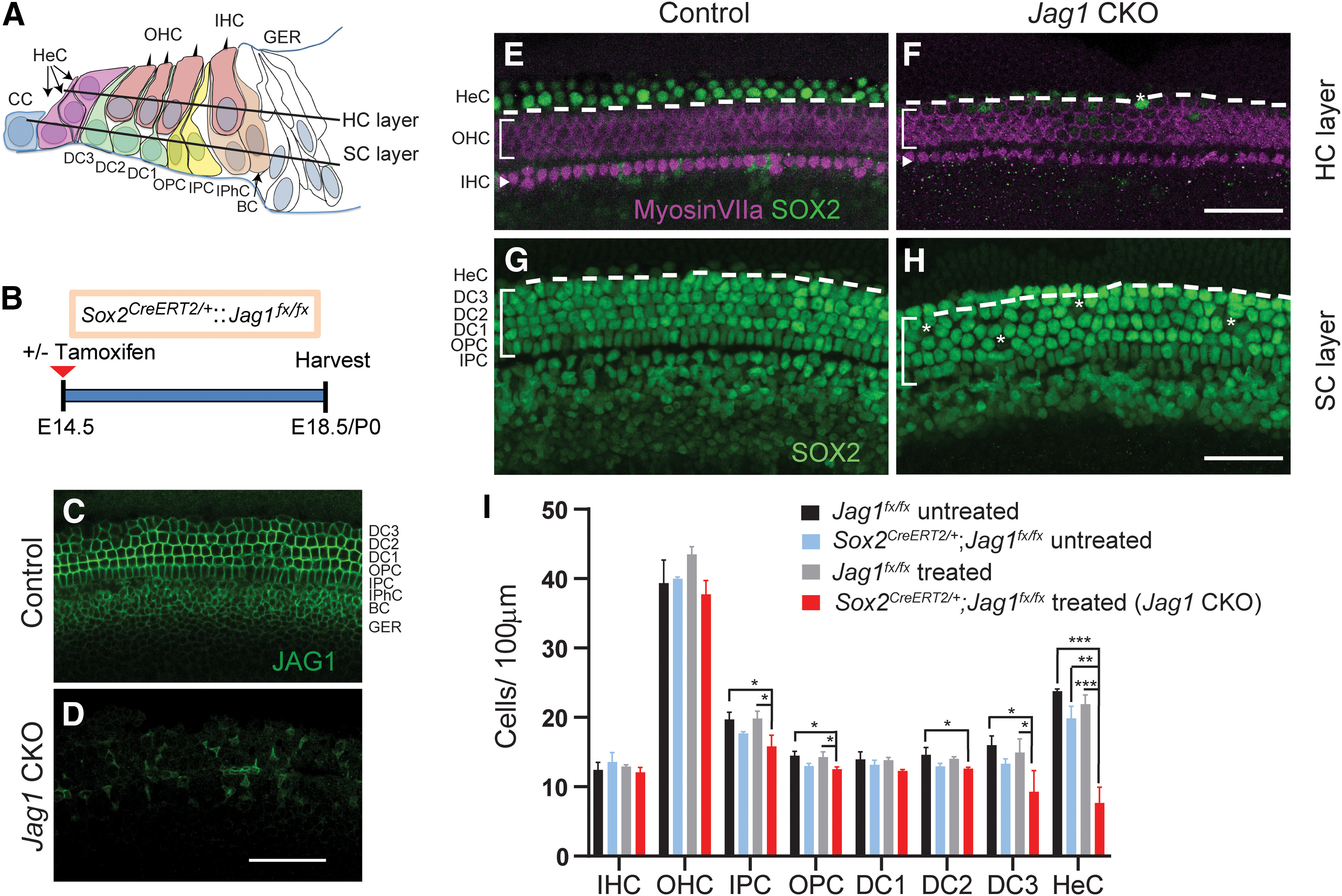
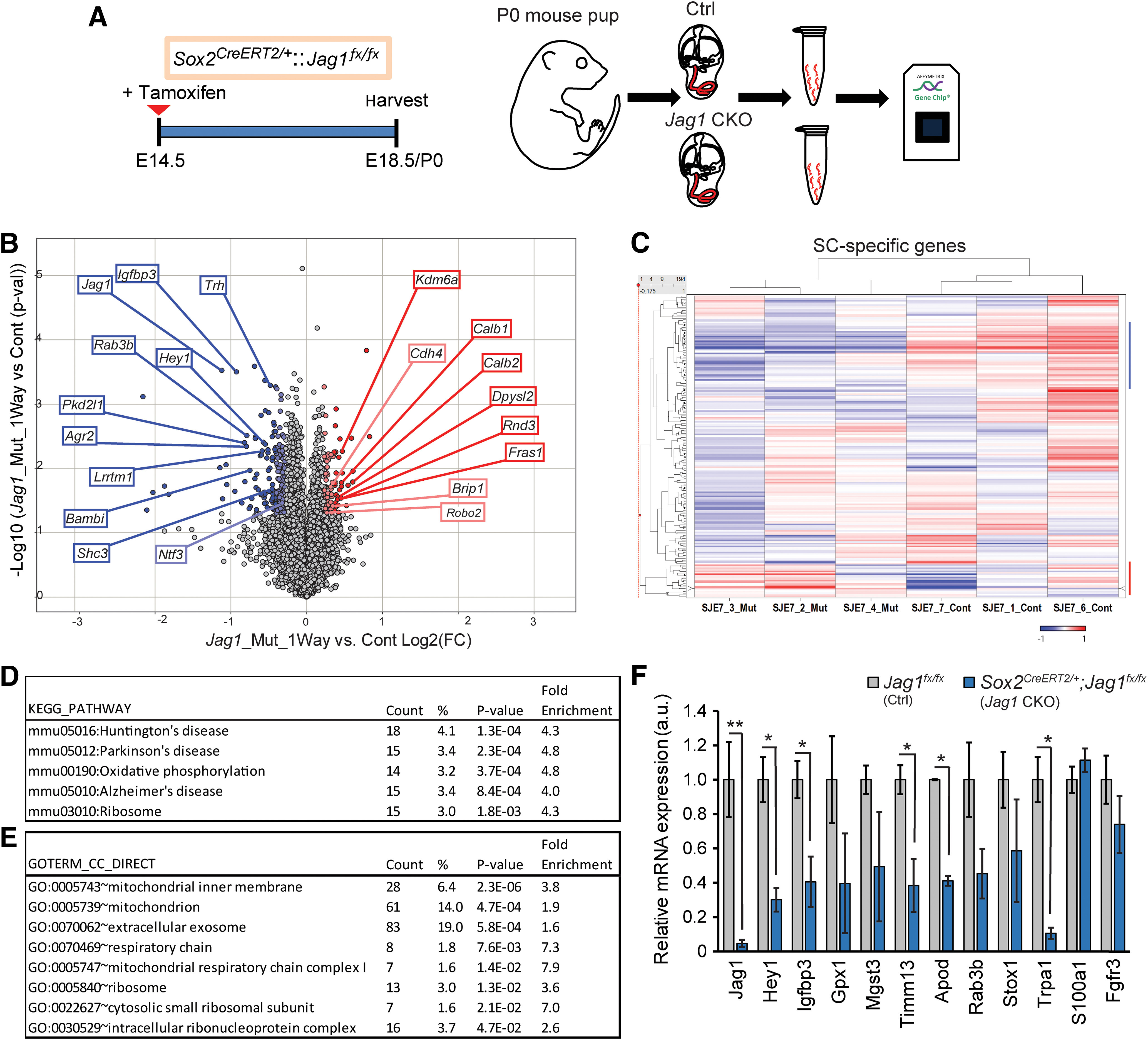
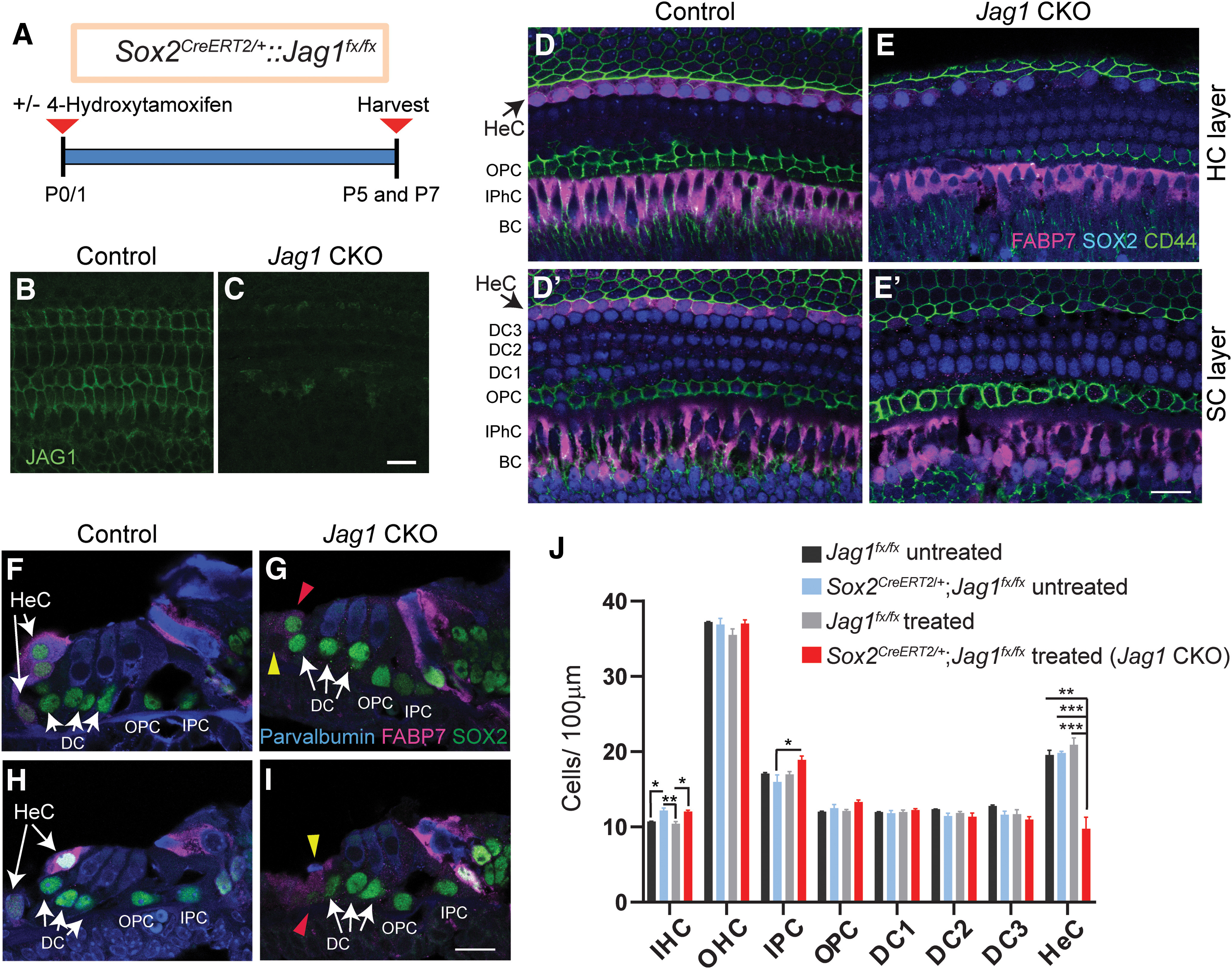
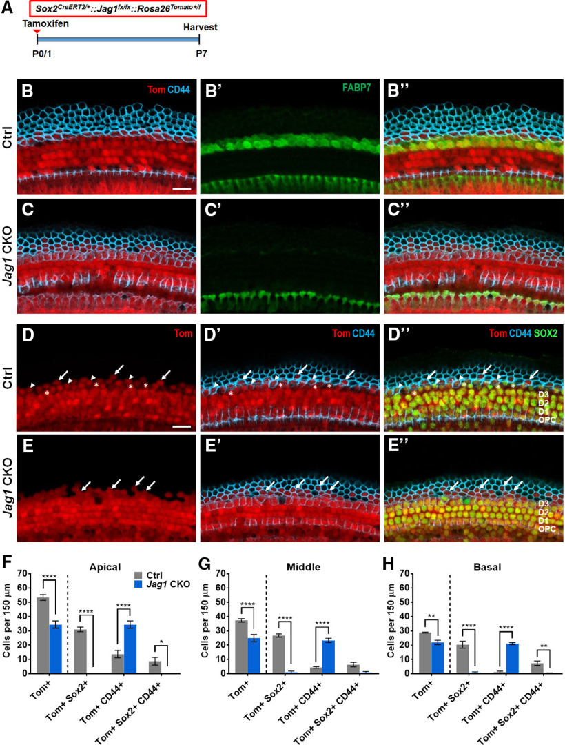
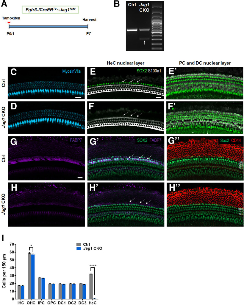
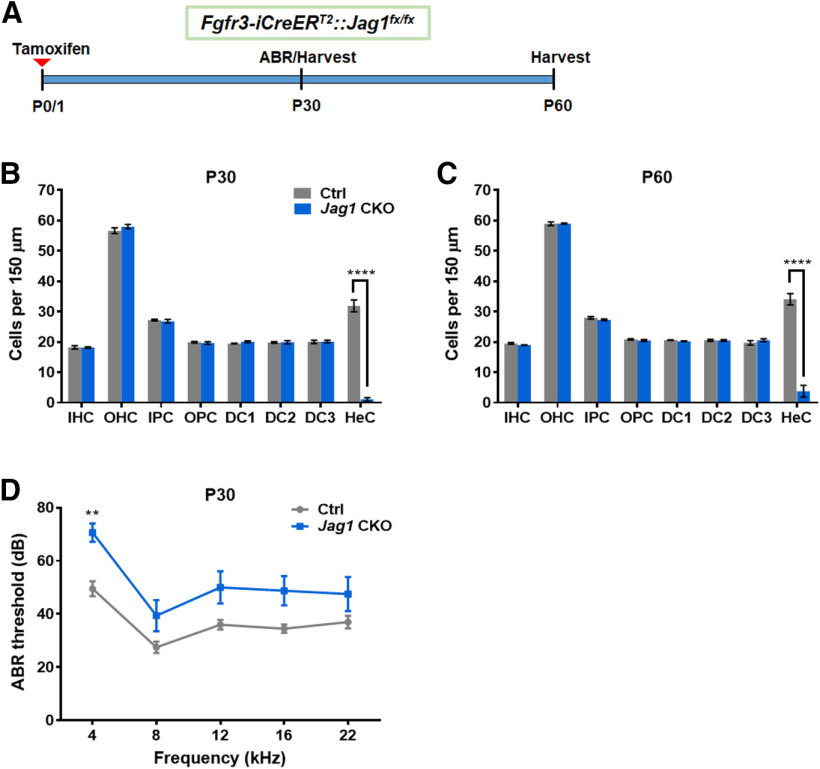
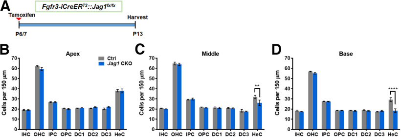
During cochlear development, the Notch ligand JAGGED 1 (JAG1) plays an important role in the specification of the prosensory region, which gives rise to sound-sensing hair cells and neighboring supporting cells (SCs). While JAG1's expression is maintained in SCs through adulthood, the function of JAG1 in SC development is unknown. Here, we demonstrate that JAG1 is essential for the formation and maintenance of Hensen's cells, a highly specialized SC subtype located at the edge of the auditory epithelium. Using Sox2CreERT2/+::Jag1loxP/loxP mice of both genders, we show that Jag1 deletion at the onset of differentiation, at embryonic day 14.5, disrupted Hensen's cell formation. Similar loss of Hensen's cells was observed when Jag1 was deleted after Hensen's cell formation at postnatal day (P) 0/P1 and fate-mapping analysis revealed that in the absence of Jag1, some Hensen's cells die, but others convert into neighboring Claudius cells. In support of a role for JAG1 in cell survival, genes involved in mitochondrial function and protein synthesis were downregulated in the sensory epithelium of P0 cochlea lacking Jag1 Finally, using Fgfr3-iCreERT2 ::Jag1loxP/loxP mice to delete Jag1 at P0, we observed a similar loss of Hensen's cells and found that adult Jag1 mutant mice have hearing deficits at the low-frequency range.SIGNIFICANCE STATEMENT Hensen's cells play an essential role in the development and homeostasis of the cochlea. Defects in the biophysical or functional properties of Hensen's cells have been linked to auditory dysfunction and hearing loss. Despite their importance, surprisingly little is known about the molecular mechanisms that guide their development. Morphologic and fate-mapping analyses in our study revealed that, in the absence of the Notch ligand JAGGED1, Hensen's cells died or converted into Claudius cells, which are specialized epithelium-like cells outside the sensory epithelium. Confirming a link between JAGGED1 and cell survival, transcriptional profiling showed that JAGGED1 maintains genes critical for mitochondrial function and tissue homeostasis. Finally, auditory phenotyping revealed that JAGGED1's function in supporting cells is necessary for low-frequency hearing.

Keywords: Claudius cells; Hensen's cells; Jagged1; Notch; cochlea development; supporting cells.


Figures

Figure 1.
Figure 1.
Deletion of Jag1 from stage E14.5 progenitor cells disrupts Hensen's cell formation. A, Schematic of the cellular composition of the neonatal auditory sensory epithelium. BC, Border cell; CC, Claudius cell; DC1, Deiters' cell row 1; DC2, Deiters' cell row 2; DC3, Deiters' cell row 3; GER, greater epithelial ridge; HeC, Hensen's cell, IHC, inner hair cell; IPC, inner pillar cell; IPhC, inner phalangeal cell; OHC, outer hair cell; OPC, outer pillar cell. B, Experimental strategy. Timed pregnant dams were injected with tamoxifen and progesterone at E14.5. Jag1 CKO (Sox2CreERT2/+::Jag1fx/fx treated) mice and control (Jag1fx/fx treated) littermates were analyzed at E18.5/P0. SOX2 immunostaining labels SC nuclei, including Hensen's cells, while myosin VIIa immunostaining labels IHCs and OHCs. Representative confocal images of the cochlear mid-turn are shown. To control for Sox2 haploinsufficiency, E18.5/P0 Sox2CreERT2/+::Jag1fx/fx mice and Jag1fx/fx littermates that did not receive tamoxifen and progesterone (untreated) were also analyzed. C, D, Validation of Jag1 deletion. SC nuclear layer immunostained for JAG1 (green), in Jag1 CKO (D) and control (C, treated, Jag1fx/fx) mice. E, F, HC and Hensen's cell phenotype in P0 control (E, Sox2CreERT2/+::Jag1fx/fx untreated) and P0 Jag1 CKO (F) mice. Control mice (E) contain 1 or 2 rows of SOX2+ (green) Hensen's cells lateral to the myosin VIIa+ (magenta) OHCs (atop dashed white line) (see schematic in A), whereas Jag1 mutant mice contain only few scattered Hensen's cells (white asterisk). G, H, SC phenotype in E18.5 control (G, Jag1fx/fx treated) and E18.5 Jag1 CKO (H) mice. SOX2+ Deiters' cell and Hensen's cell nuclei (atop dashed white line) (see schematic in A) are enlarged and misaligned (white asterisks) in Jag1 CKO mice. I, Quantification of HC and SC subtypes in control and Jag1 CKO mice. Data are mean ± SD; n = 3/group. *p < 0.05; **p ≤ 0.001; ***p ≤ 0.0001; two-way ANOVA, with Bonferroni correction. Scale bar, 50 μm.
Figure 2.
Figure 2.
Loss of JAG1 leads to the downregulation of genes involved in mitochondrial function and protein synthesis. A, Experimental strategy. Timed pregnant dams were injected with tamoxifen and progesterone at E14.5, and gene expression in cochlear epithelia from E18.5/P0 Jag1 CKO, (Sox2CreERT2/+::Jag1fx/fx) mice, and control (Ctrl, Jag1fx/fx) littermates was analyzed using Clariom D mouse arrays. B, Volcano plot presents the one-way ANOVA of differential gene expression in Jag1 CKO versus control samples (n = 3 animals/group). Plotted as log2 fold-change (FC) (x axis) versus −log10 p value (y axis). Dark blue (log2 (FC) ≤ −6σ) and light blue (log2 (FC) ≤ −3σ) dots represent downregulated genes. Dark red (log2 (FC) ≥ 6σ) and light red dots (log2 (FC) ≥ 3σ) represent upregulated genes. C, Heat cluster map illustrating JAG1-mediated effects on a list of 200 SC-specific genes curated from published data. D, E, GO analysis of genes downregulated (mean log2 control ≤ 5, p ≤ 0.05, log2 (FC) ≤ −2σ) in P0 Jag1 CKO versus control (Ctrl) cochlear epithelia. Listed are significantly enriched pathways (D) and cellular component (E) GO terms. p values were calculated using Benjamini correction. F, qRT-PCR-based validation of genes of interest that were downregulated at P0 in Jag1 CKO cochlear epithelia compared with Ctrl. SC-specific marker genes S100a1 and Fgfr3 served as controls and no difference between groups was seen. Data are mean ± SEM; minimum of n = 3 animals/group. *p < 0.05; **p ≤0.001; two-tailed Student's t test.
Figure 3.
Figure 3.
Deletion of Jag1 from neonatal SCs results in the loss of Hensen's cells. A, Experimental strategy. Sox2CreERT2/+::Jag1fx/fx and Jag1fx/fx were treated with 4-hydroxy-tamoxifen at P0/P1, and their cochleae were analyzed at P5 or P7. Untreated control mice (Sox2CreERT2/+::Jag1fx/fx untreated and Jag1fx/fx, untreated) were also used. B, C, Validation of conditional Jag1 deletion. Shown is the SC nuclear layer, immunostained for JAG1 (green), in Jag1 CKO (C) and control (B, Jag1fx/fx treated) mice. D–I, SOX2 immunostaining (D–E′, blue; F–I, green) labels SC nuclei, including Hensen's cells. FABP7 (magenta) labels Hensen's cells, inner phalangeal cells, and border cells. Parvalbumin (F–I, blue) labels HCs, and CD44 (D–E′, green) labels Claudius cells and outer pillar cells. Shown are representative single-plane confocal images of the cochlear mid-turn. D–E′, Top down view of the HC (D,E) and SC nuclear layers (D′,E′) in Jag1 CKO (E,E′) and control (D,D′, Sox2CreERT2/+::Jag1fx/fx untreated) mice. Black arrows indicate the location of Hensen's cells residing within the HC (D) and SC layer (D′) in control mice. F–I, Cochlear cross-sections of Jag1 CKO (G,I) and control mice (F,H, Sox2CreERT2/+::Jag1fx/fx, untreated). White arrows indicate 2 or 3 Hensen's cell nuclei that are stacked on top of each other in control mice. Red arrowheads indicate SOX2+ Hensen's cell-like cells (G,I). Yellow arrowheads indicate dying cell (I) and missing Hensen's cell (G) in Jag1 CKO mice. J, Quantification of HC and SC subtypes in control and Jag1 CKO mice. Data are mean ± SD; n = 3/group. *p < 0.05; **p ≤ 0.001; ***p ≤ 0.0001; two-way ANOVA, with Bonferroni correction. DC1, Deiters' cell row 1; DC2, Deiters' cell row 2; DC3, Deiters' cell row 3; HeC, Hensen's cell; IPC, inner pillar cell; IPhC, inner phalageal cell; OPC, outer pillar cell. Scale bar, 20 μm.
Figure 4.
Figure 4.
Fate-mapping analysis reveals that Hensen's cells die or convert into Claudius cells after deletion of Jag1. A, Experimental strategy. Sox2CreERT2/+::Jag1fx/fx::Rosa26tdTomato/+ (Jag1 CKO) and Sox2CreERT2/+::Rosa26tdTomato/+ (Ctrl) mice were injected with tamoxifen at P0/P1, and cochleae were analyzed at P7. B–C′′, Representative confocal images from Ctrl (B–B′′) and Jag1 CKO (C–C′′) mice in the apical turn, showing expression of FABP7 (a Hensen's cell marker, green), CD44 (a Claudius cell marker, blue), and tdTomato (Tom, red). D–E′′, Representative confocal images of Ctrl (D–D′′) and Jag1 CKO (E–E′′) mice, showing expression of SOX2 (a SC marker, green), CD44 (blue), and Tom (red). Arrows indicate Tom+, CD44+ cells. Asterisks indicate Tom+, SOX2+ cells. Arrowheads indicate Tom+, SOX2+, CD44+ cells. F–H, Quantification of Tom+ cells lateral to the third row of Deiters' cells that also express CD44 and/or SOX2 in the apical (F), middle (G), and basal (H) turns of the cochleae in Ctrl and Jag1 CKO mice. Data are mean ± SEM; n = 3 or 4/group. *p < 0.05; **p < 0.01; ****p < 0.0001; two-way ANOVA followed by a Bonferroni post hoc test. Scale bar, 20 μm.
Figure 5.
Figure 5.
Massive loss of Hensen's cells at P7 after neonatal deletion of Jag1 from pillar and Deiters' cells. A, Experimental strategy. Fgfr3-iCreERT2::Jag1fx/fx (Jag1 CKO) and Jag1fx/fx (Ctrl) mice were injected with tamoxifen at P0/P1, and cochleae were analyzed at P7. B, RT-PCR results using RNA extracted from P7 Ctrl and Jag1 CKO whole cochlea. The upper band (541 bp) indicates the WT allele containing exon 4. The lower band (286 bp, white arrow) confirms deletion of Jag1 exon 4 in Jag1 CKO mice. The upper band is still present in Jag1 CKO mice since JAG1 is expressed in all SCs and Fgfr3-iCreERT2 only targets pillar and Deiters' cells. C, D, Representative confocal slice images of HC, labeled with myosin VIIa (blue) in P7 Ctrl (C) and Jag1 CKO (D) mice. E–F′, Representative confocal slice images of SC nuclear layers at the level of Hensen's cell nuclei (E,F) and at the level of pillar and Deiters' cell nuclei (E′,F′) showing SOX2 (an SC marker, green) and S100a1 (a pillar and Deiters' cell marker, white) expression in P7 Ctrl (E–E′) and Jag1 CKO (F–F′) mice. Arrows indicate the location of Hensen's cells (E,F). G–H′′, Representative confocal slice images of SOX2 (green), FABP7 (a Hensen's cell marker, magenta), and CD44 (a Claudius cell marker, red) expression in P7 Ctrl (G–G′′) and Jag1 CKO (H–H′′) mice. Arrows indicate the location of Hensen's cells (G′,H′). I, Quantification of IHCs, OHCs, and individual SC subtypes (inner pillar cells to Hensen's cells) in Ctrl and Jag1 CKO mice. Data are mean ± SEM; n = 4/group. *p < 0.05; ****p < 0.0001; two-way ANOVA followed by a Bonferroni post hoc test. DC1, Deiters' cell row 1; DC2, Deiters' cell row 2; DC3, Deiters' cell row 3; HeC, Hensen's cell; IPC, inner pillar cell; OPC, outer pillar cell. Scale bar, 20 μm.
Figure 6.
Figure 6.
Neonatal Jag1 deletion from pillar and Deiters' cells caused hearing deficits in the low-frequency range. A, Experimental strategy. Fgfr3-iCreERT2::Jag1fx/fx (Jag1 CKO) and Jag1fx/fx (Ctrl) mice were injected with tamoxifen at P0/P1, followed by ABR at P30, and analysis of the cochlea at P30 or P60. B, C, Quantification of IHCs, OHCs, and individual SC subtypes (inner pillar cells to Hensen's cells) in Ctrl and Jag1 CKO mice at P30 (B) and P60 (C). Data are mean ± SEM; n = 4/group. ****p < 0.0001. D, ABR thresholds of Ctrl and Jag1 CKO at P30. Data are mean ± SEM; n = 8-10/group. **p < 0.01; two-way ANOVA followed by a Bonferroni post hoc test. DC1, Deiters' cell row 1; DC2, Deiters' cell row 2; DC3, Deiters' cell row 3; HeC, Hensen's cell; IPC, inner pillar cells; OPC, outer pillar cells.
Figure 7.
Figure 7.
Mild loss of Hensen's cells after deletion of Jag1 from pillar and Deiters' cells at 1 week of age. A, Experimental strategy. Fgfr3-iCreERT2::Jag1fx/fx (Jag1 CKO) and Jag1fx/fx (Ctrl) mice were injected with tamoxifen at P6/P7, and cochleae are analyzed at P13. B–D, Quantification of IHCs, OHCs, and individual SC subtypes (inner pillar cells to Hensen's cells) in the apical (B), middle (C), and basal (D) turns of the cochleae in Ctrl and Jag1 CKO mice at P13. Data are mean ± SEM; n = 4/group. **p < 0.01; ****p < 0.0001; two-way ANOVA followed by a Bonferroni post hoc test. DC1, Deiters' cell row 1; DC2, Deiters' cell row 2; DC3, Deiters' cell row 3; HeC, Hensen's cell; IPC, inner pillar cells; OPC, outer pillar cells.

References

    1. Adam J, Myat A, Le Roux I, Eddison M, Henrique D, Ish-Horowicz D, Lewis J (1998) Cell fate choices and the expression of Notch, Delta and Serrate homologues in the chick inner ear: parallels with Drosophila sense-organ development. Development 125:4645–4654. - PubMed
    1. Androutsellis-Theotokis A, Leker RR, Soldner F, Hoeppner DJ, Ravin R, Poser SW, Rueger MA, Bae SK, Kittappa R, McKay RD (2006) Notch signalling regulates stem cell numbers in vitro and in vivo. Nature 442:823–826. 10.1038/nature04940 - DOI - PubMed
    1. Arnold K, Sarkar A, Yram MA, Polo JM, Bronson R, Sengupta S, Seandel M, Geijsen N, Hochedlinger K (2011) Sox2(+) adult stem and progenitor cells are important for tissue regeneration and survival of mice. Cell Stem Cell 9:317–329. 10.1016/j.stem.2011.09.001 - DOI - PMC - PubMed
    1. Atkinson PJ, Dong Y, Gu S, Liu W, Najarro EH, Udagawa T, Cheng AG (2018) Sox2 haploinsufficiency primes regeneration and Wnt responsiveness in the mouse cochlea. J Clin Invest 128:1641–1656. 10.1172/JCI97248 - DOI - PMC - PubMed
    1. Basch ML, Ohyama T, Segil N, Groves AK (2011) Canonical Notch signaling is not necessary for prosensory induction in the mouse cochlea: insights from a conditional mutant of RBPjκ. J Neurosci 31:8046–8058. 10.1523/JNEUROSCI.6671-10.2011 - DOI - PMC - PubMed

Publication types