

Macular vessel density and foveal avascular zone parameters in patients after acute primary angle closure determined by OCT angiography
- PMID: 33127916
- PMCID: PMC7603312
- DOI: 10.1038/s41598-020-73223-9
Macular vessel density and foveal avascular zone parameters in patients after acute primary angle closure determined by OCT angiography
Abstract
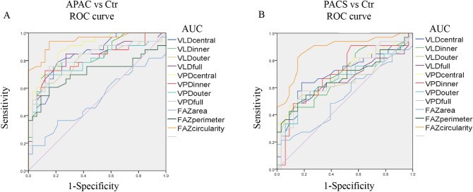
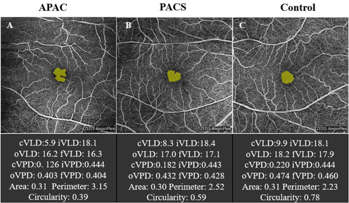
This study analyzed the optical coherence tomography angiography (OCTA) macular parameters in primary angle-closure glaucoma (PACG) patients after acute primary angle closure (APAC) episodes. Thirty-three patients with 33 APAC eyes and 33 primary angle closure suspect (PACS) eyes and 33 age-matched normal subjects (controls) were enrolled. Macular vessel density (VD) in central, inner, outer and full regions and foveal avascular zone (FAZ) parameters (area, perimeter and circularity index) were compared between APAC, PACS, and control eyes. For resolved APAC eyes, the VD in each macular region was significantly lower than that in control eyes, with less central and inner macular VD than PACS eyes. The central macular VD was significantly lower in PACS eyes than in controls. There was no difference in FAZ area and perimeter between APAC, PACS, and control eyes. FAZ circularity was highest in control eyes, followed by PACS eyes, and lowest in APAC eyes. The AUC, sensitivity and specificity of FAZ circularity were 0.944, 93.9% and 84.8%, respectively, in APAC eyes and 0.881, 84.8% and 81.8%, respectively, in PACS eyes. Therefore, FAZ circularity had the best discrimination capability for detecting both APAC and PACS eyes. Macular assessment with OCTA could provide an accurate early-stage diagnostic tool for PACG.
Conflict of interest statement
The authors declare no competing interests.
Figures
Similar articles
-
Quantitative optical coherence tomography angiography of macular vascular structure and foveal avascular zone in glaucoma.PLoS One. 2017 Sep 21;12(9):e0184948. doi: 10.1371/journal.pone.0184948. eCollection 2017. PLoS One. 2017. PMID: 28934255 Free PMC article.
-
Changes of Optic Disc and Macular Vessel Perfusion Density in Primary Angle Closure Glaucoma: A Quantitative Study Using Optical Coherence Tomography Angiograph.Ophthalmic Res. 2023;66(1):1245-1253. doi: 10.1159/000533874. Epub 2023 Aug 30. Ophthalmic Res. 2023. PMID: 37647877 Free PMC article.
-
[Correlation of capillary plexus with visual acuity in idiopathic macular epiretinal membrane eyes using optical coherence tomography angiography].Zhonghua Yan Ke Za Zhi. 2019 Oct 11;55(10):757-762. doi: 10.3760/cma.j.issn.0412-4081.2019.10.006. Zhonghua Yan Ke Za Zhi. 2019. PMID: 31607064 Chinese.
-
Retinal microvasculature features in patients with Behcet's disease: a systematic review and meta-analysis.Sci Rep. 2022 Jan 14;12(1):752. doi: 10.1038/s41598-021-04730-6. Sci Rep. 2022. PMID: 35031636 Free PMC article.
-
Application of Optical Coherence Tomography Angiography Macular Analysis for Systemic Hypertension. A Systematic Review and Meta-analysis.Am J Hypertens. 2022 Apr 2;35(4):356-364. doi: 10.1093/ajh/hpab172. Am J Hypertens. 2022. PMID: 34718393
Cited by
-
Quantitative Measurements of Vessel Density and Blood Flow Areas Primary Angle Closure Diseases: A Study of Optical Coherence Tomography Angiography.J Clin Med. 2022 Jul 13;11(14):4040. doi: 10.3390/jcm11144040. J Clin Med. 2022. PMID: 35887804 Free PMC article.
-
Body shape and risk of glaucoma: A Mendelian randomization.Front Med (Lausanne). 2022 Sep 23;9:999974. doi: 10.3389/fmed.2022.999974. eCollection 2022. Front Med (Lausanne). 2022. PMID: 36213644 Free PMC article.
-
Alterations in Macular Microvasculature in Pterygium Patients Measured by OCT Angiography.Diagnostics (Basel). 2023 Apr 30;13(9):1603. doi: 10.3390/diagnostics13091603. Diagnostics (Basel). 2023. PMID: 37174994 Free PMC article.
-
Spatial positional relationship between macular superficial vessel density and ganglion cell-inner plexiform layer thickness in primary angle closure glaucoma.Int Ophthalmol. 2022 Jan;42(1):103-112. doi: 10.1007/s10792-021-02005-7. Epub 2021 Aug 15. Int Ophthalmol. 2022. PMID: 34392472 Free PMC article.
-
Macular vascular density changes in different stages of chronic primary angle-closure glaucoma.Front Med (Lausanne). 2025 Jul 24;12:1620673. doi: 10.3389/fmed.2025.1620673. eCollection 2025. Front Med (Lausanne). 2025. PMID: 40776918 Free PMC article.
References
Publication types
MeSH terms
Substances
LinkOut - more resources
Full Text Sources
