

Anterior chamber angle imaging with swept-source optical coherence tomography: comparison between CASIAII and ANTERION
- PMID: 33127944
- PMCID: PMC7603499
- DOI: 10.1038/s41598-020-74813-3
Anterior chamber angle imaging with swept-source optical coherence tomography: comparison between CASIAII and ANTERION
Abstract
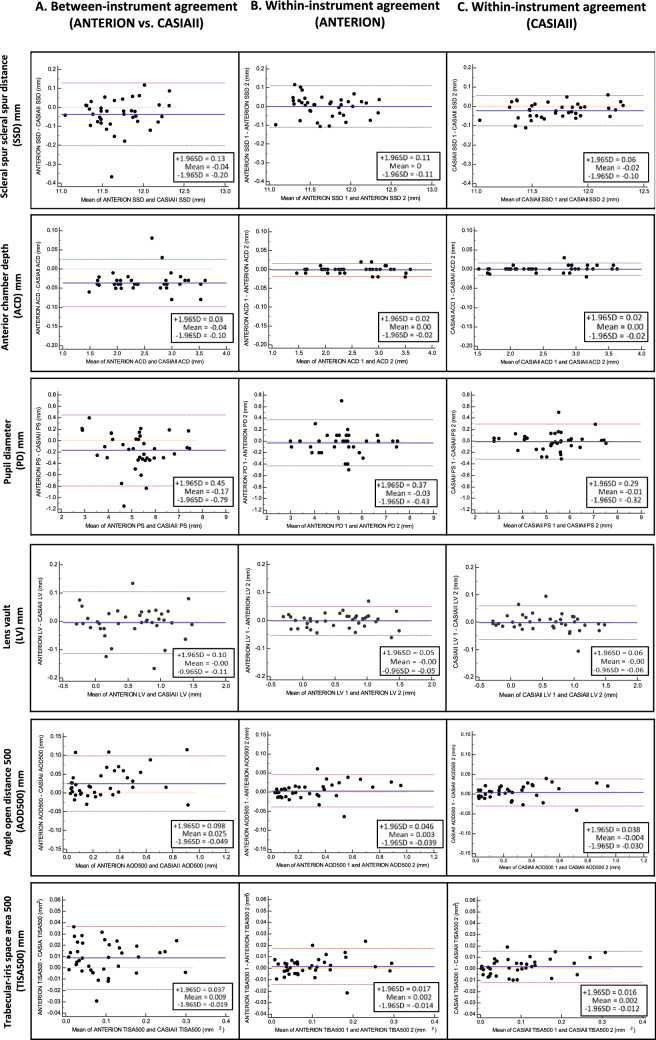
This study compared the test-retest variabilities and measurement agreement of anterior chamber angle (ACA) dimensions measured by two anterior segment swept-source optical coherence tomography (SS-OCT)-the ANTERION (Heidelberg Engineering, Heidelberg, Germany) and CASIAII (Tomey, Nagoya, Japan). Thirty-eight subjects, 18 patients with primary angle closure and 20 healthy participants with open angles, were included. The mean age was 54.7 ± 15.8 years (range: 26-75 years). One eye of each subject was randomly selected for anterior segment imaging by ANTERION and CASIAII, using the same scan pattern (6 evenly spaced radial scans across the anterior segment for three times) in the same visit. The between- and within-instrument agreement and repeatability coefficients of angle open distance (AOD500), trabecular-iris space area (TISA500), lens vault (LV), scleral spur-scleral spur distance (SSD), anterior chamber depth (ACD), and pupil diameter (PD) were measured. The anterior and posterior boundaries of the cornea, iris, and lens were automatically segmented by the SS-OCT instruments; the scleral spur was manually located by a single masked observer. There were significant differences between ANTERION and CASIAII measurements; the SSD, PD, and ACD were smaller whereas AOD500 and TISA500 were greater in ANTERION compared with CASIAII (P < 0.001). Anterior segment measurements obtained from the two SS-OCT instruments showed strong associations (R2 ranged between 0.866 and 0.998) although the between-instrument agreement was poor; the spans of 95% limits of between-instrument agreement were ≥ 1.5-folds than the within-instrument agreement for either instrument. Whereas both SS-OCT instruments showed low test-retest measurement variabilities, the repeatability coefficients of AOD500, TISA500, ACD, and PD were slightly smaller for CASIAII than ANTERION (P ≤ 0.012).
Conflict of interest statement
CL has received research support in the form of instrument and speaker honorarium from Tomey (Japan) and Heidelberg Engineering (Germany). Other authors do not have any other competing interests.
Figures


References
Publication types
MeSH terms
LinkOut - more resources
Full Text Sources
