. 2020 Oct 30;10(1):18719.
doi: 10.1038/s41598-020-75761-8.

Mild electrical stimulation with heat shock attenuates renal pathology in adriamycin-induced nephrotic syndrome mouse model

Affiliations

Mild electrical stimulation with heat shock attenuates renal pathology in adriamycin-induced nephrotic syndrome mouse model

Keisuke Teramoto et al. Sci Rep. .

Abstract

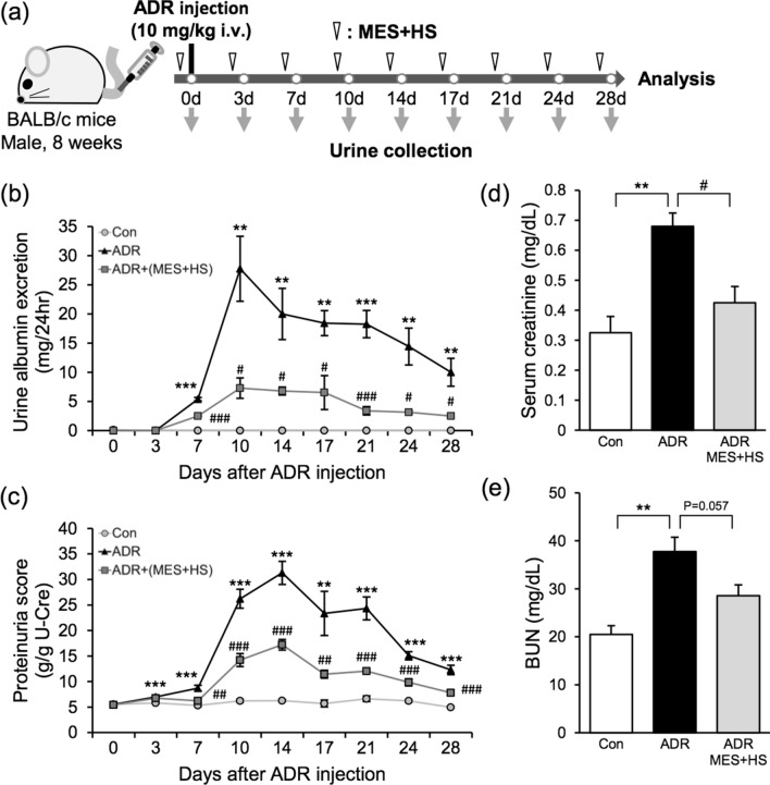
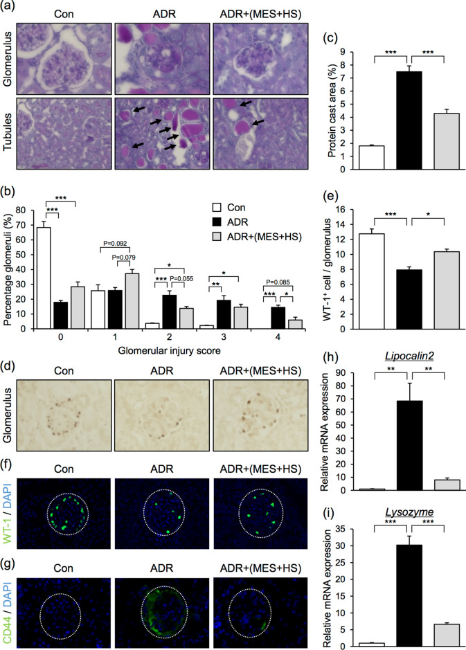
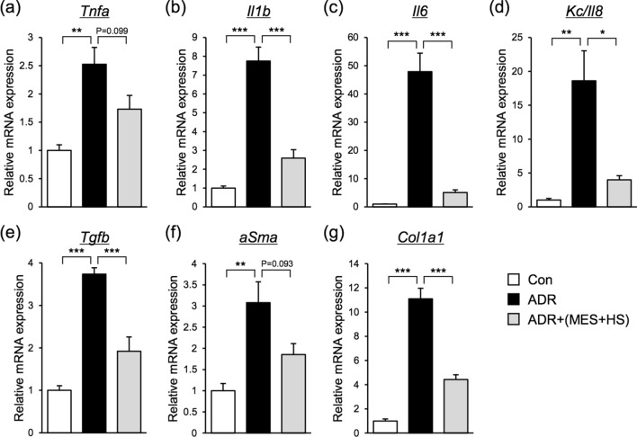
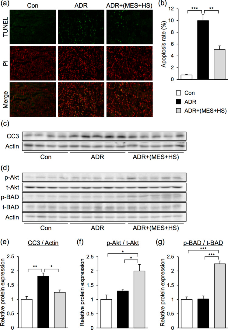
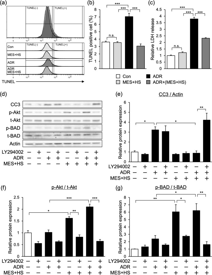
Nephrotic syndrome (NS) is a renal disorder that is characterized by massive proteinuria, hypoalbuminemia and edema. One of the main causes of NS is focal segmental glomerulosclerosis (FSGS), which has extremely poor prognosis. Although steroids and immunosuppressants are the first line of treatment, some FSGS cases are refractory, prompting the need to find new therapeutic strategies. We have previously demonstrated that an optimized combination treatment of mild electrical stimulation (MES) and heat shock (HS) has several biological benefits including the amelioration of the pathologies of the genetic renal disorder Alport syndrome. Here, we investigated the effect of MES + HS on adriamycin (ADR)-induced NS mouse model. MES + HS suppressed proteinuria and glomerulosclerosis induced by ADR. The expressions of pro-inflammatory cytokines and pro-fibrotic genes were also significantly downregulated by MES + HS. MES + HS decreased the expression level of cleaved caspase-3 and the number of TUNEL-positive cells, indicating that MES + HS exerted anti-apoptotic effect. Moreover, MES + HS activated the Akt signaling and induced the phosphorylation and inhibition of the apoptotic molecule BAD. In in vitro experiment, the Akt inhibitor abolished the MES + HS-induced Akt-BAD signaling and anti-apoptotic effect in ADR-treated cells. Collectively, our study suggested that MES + HS modulates ADR-induced pathologies and has renoprotective effect against ADR-induced NS via regulation of Akt-BAD axis.


Conflict of interest statement

The authors declare no competing interests.

Figures

Figure 1
Figure 1
MES + HS ameliorates renal dysfunction in ADR-induced NS mice. (a) Experimental diagram originally drawn by us. Eight-week-old male mice were injected with ADR (10 mg/kg; i.v.) and treated with MES + HS for 10 min twice a week for 4 weeks. MES + HS treatment was started one day prior to injection with ADR. Urine samples were collected twice a week. (b) Urinary albumin excretion was assessed by 12% SDS-PAGE followed by CBB staining and densitometric analysis of gel blots. (c) Urinary protein and creatinine were assessed by Bradford’s and Jaffe’s method, respectively. Urinary protein concentration was normalized with urinary creatinine concentration. (d) Serum creatinine and (e) blood urea nitrogen (BUN) scores of murine plasma was measured using DRI-CHEM (Fuji). Values are the mean ± SE. n = 4–5 mice per group. **p < 0.01, ***p < 0.001 (Con vs ADR), #p < 0.05, ##p < 0.01, ###p < 0.001 [ADR vs ADR + (MES + HS)] assessed by Tukey–Kramer method.
Figure 2
Figure 2
MES + HS improves the renal pathology in ADR-induced NS mouse model. (a) Images of PAS-stained kidney section. Upper images show glomerulus and lower images show tubules. Arrows indicate protein casts. (b,c) Bar graph shows quantification of glomerulosclerosis severity score and protein cast area, respectively. (d) Glomerular sections were immunostained to detect WT-1. (e) Bar graph shows the ratio of WT-1-positive cells to glomerulus. (f,g) Glomerular sections were stained with WT-1 and CD44, respectively, and visualized by immunofluorescence. (h,i) Total RNA was isolated from renal tissues and assessed by quantitative RT-PCR analysis. Bar graphs show gene expression levels of renal injury markers (Lipocalin2, Lysozyme). Values were normalized to 18S ribosomal RNA (internal control). Values are the mean ± SE. n = 4–5 mice per group. *p < 0.05, **p < 0.01, ***p < 0.001 assessed by Tukey–Kramer method.
Figure 3
Figure 3
MES + HS suppresses the expression levels of pro-inflammatory cytokines and pro-fibrotic genes. (ag) Total RNA was isolated from renal tissues, and pro-inflammatory (ad) and pro-fibrotic genes (eg) were assessed by quantitative RT-PCR. Values were normalized to 18S ribosomal RNA (internal control). Values are the mean ± SE. n = 4–5 mice per group. *p < 0.05, **p < 0.01, ***p < 0.001 assessed by Tukey–Kramer method.
Figure 4
Figure 4
MES + HS suppresses apoptosis in ADR-induced NS mice. (a) Renal sections were stained with TUNEL for apoptotic cells. All cells were counterstained with PI. (b) Apoptosis rate was calculated by the ratio of TUNEL-positive cells to PI-positive cells. (c,d) Immunoblots of lysates isolated from mouse renal tissues were analyzed by western blotting using the indicated antibodies. Actin was used as loading control. Full-length blots are presented in Supplementary Figure 1. (eg) Bar graph shows quantification of blots in (c,d). Expression of phosphorylated proteins were normalized with total protein expression. Graphs are the mean ± SE. n = 4–5 mice per group. *p < 0.05, **p < 0.01, ***p < 0.001 assessed by Tukey–Kramer method.
Figure 5
Figure 5
MES + HS suppresses ADR-induced apoptosis in MPC5 cell line. (a) MPC5 cells were treated with MES + HS (1 V/cm, 0.1 ms, 55 pps) for 10 min, and subsequently treated with 200 nmol/L ADR. Twenty-four hours after, cells were stained with TUNEL and analyzed by flow cytometry. (b) Data in (a) was statistically calculated and analyzed. (c) Relative LDH release was measured by colorimetric method in MPC cells treated with MES + HS and ADR. Released LDH concentration was normalized with intracellular LDH concentration. (d) MPC5 cells were treated with Akt phosphorylation inhibitor (LY294002; 10 µM) for 1 h before MES + HS and ADR (200 nmol/L) treatment. Protein lysates were collected 24 h after ADR treatment and analyzed by western blotting. Actin was used as loading control. Full-length blots are presented in Supplementary Figure 2. (eg) Bar graph shows quantification of blots in (d). Expression of phosphorylated proteins were normalized with total protein expression. Values are the mean ± SE. n = 3 per group. *p < 0.05, **p < 0.01, ***p < 0.001 assessed by Tukey–Kramer method.

References

    1. Mace C, Chugh SS. Nephrotic syndrome: components, connections, and angiopoietin-like 4-related therapeutics. J. Am. Soc. Nephrol. 2014;25:2393–2398. doi: 10.1681/ASN.2014030267. - DOI - PMC - PubMed
    1. Trautmann A, et al. Spectrum of steroid-resistant and congenital nephrotic syndrome in children: the podoNet registry cohort. Clin. J. Am. Soc. Nephrol. 2015;10:592–600. doi: 10.2215/CJN.06260614. - DOI - PMC - PubMed
    1. Rosenberg AZ, Kopp JB. Focal segmental glomerulosclerosis. Clin. J. Am. Soc. Nephrol. 2017;12:502–517. doi: 10.2215/CJN.05960616. - DOI - PMC - PubMed
    1. Ponticelli C, Glassock RJ. Glomerular diseases: membranous nephropathy—a modern view. Clin. J. Am. Soc. Nephrol. 2014;9:609–616. doi: 10.2215/CJN.04160413. - DOI - PMC - PubMed
    1. Bose B, Cattran D, Toronto Glomerulonephritis Registry Glomerular diseases: FSGS. Clin. J. Am. Soc. Nephrol. 2014;9:626–632. doi: 10.2215/CJN.05810513. - DOI - PMC - PubMed

Publication types

MeSH terms