. 2020 Nov;8(21):e14589.
doi: 10.14814/phy2.14589.

The NLRP3 inflammasome in macrophages is stimulated by cell-free hemoglobin

Affiliations

The NLRP3 inflammasome in macrophages is stimulated by cell-free hemoglobin

Ciara M Shaver et al. Physiol Rep. 2020 Nov.

Abstract

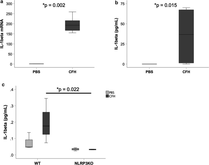
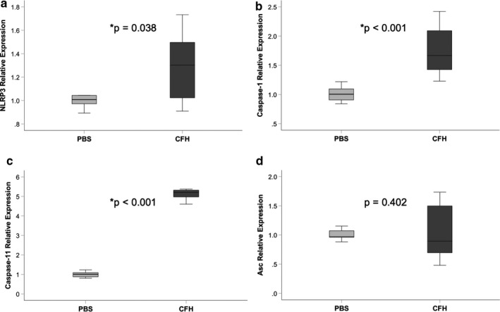
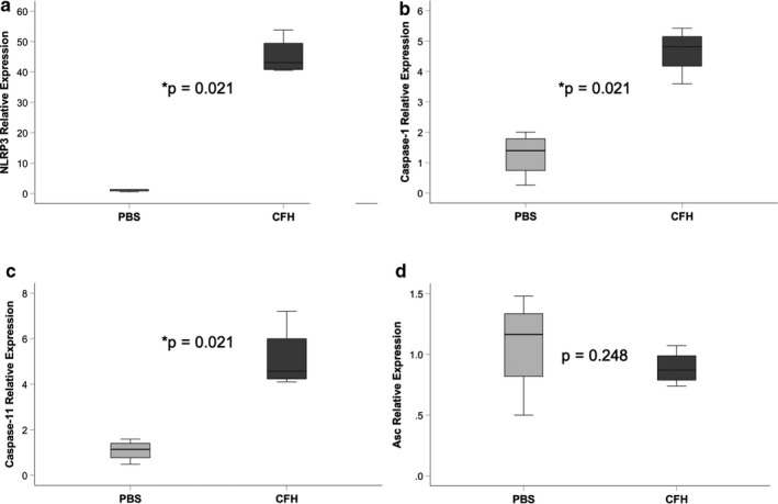
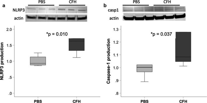
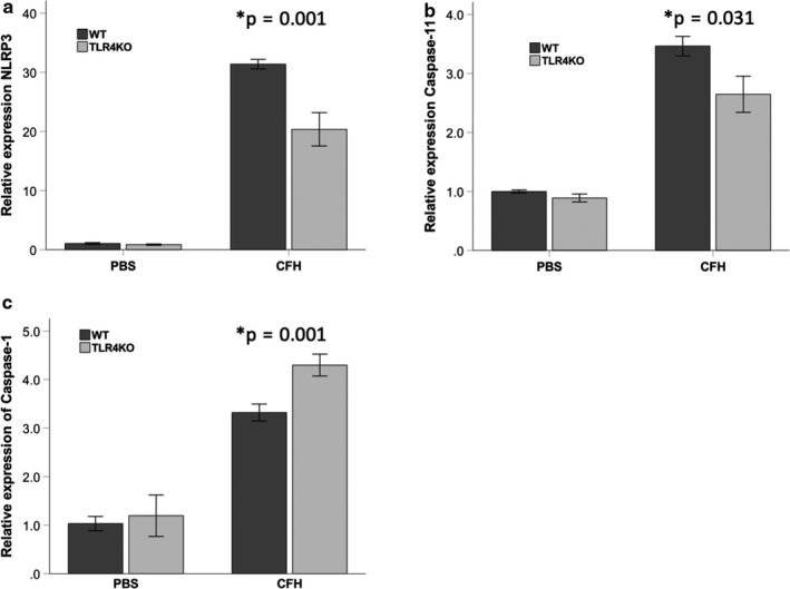
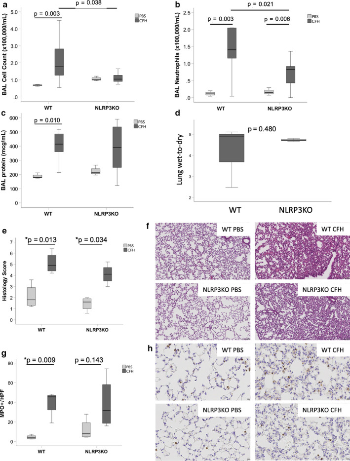
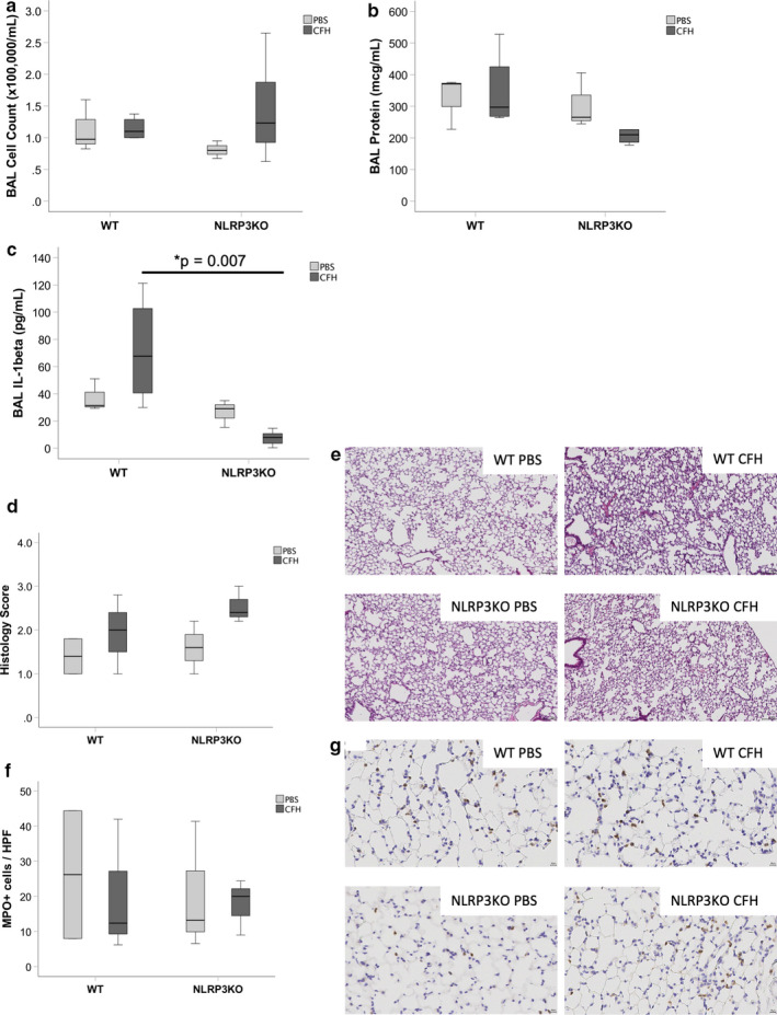
Cell-free hemoglobin (CFH) is associated with severe lung injury in human patients and is sufficient to induce airspace inflammation and alveolar-capillary barrier dysfunction in an experimental model of acute lung injury. The mechanisms through which this occurs are unknown. One key pathway which regulates inflammation during acute lung injury is the NLRP3 inflammasome. Because CFH can act as a damage-associated molecular pattern, we hypothesized that CFH may activate the NLRP3 inflammasome during acute lung injury. Primary mouse alveolar macrophages and cultured murine macrophages exposed to CFH (0-1 mg/ml) for 24 hr demonstrated robust upregulation of the NLRP3 inflammasome components NLRP3, caspase-1, and caspase-11. Maximal induction of the NLRP3 inflammasome by CFH required TLR4. Compared to wild-type controls, mice lacking NLRP3 developed less airspace inflammation (2.7 × 10⁵ cells/ml in bronchoalveolar lavage fluid versus. 1.1 × 10⁵ /ml, p = .006) after exposure to intratracheal CFH. Together, these data demonstrate that CFH can stimulate the NLRP3 inflammasome in macrophages and that this pathway may be important in the pathogenesis of CFH-induced acute lung injury.

Keywords: ARDS; NLRP3; acute lung injury; cell-free hemoglobin; inflammasome.


Conflict of interest statement

The author(s) declare no conflicts of interest.

Figures

FIGURE 1
FIGURE 1
Cell‐free hemoglobin increases expression of IL‐1β in macrophages. Cultured alveolar‐like MH‐S cells were incubated with cell‐free hemoglobin (CFH, 1 mg/ml) or PBS control for 24 hr and IL‐1β expression was measured by (a) RT‐PCR, normalized to mean expression in PBS cells and (b) ELISA. (c) Primary alveolar macrophages from wild‐type (WT) or NLRP3KO mice were incubated with CFH for 24 hr ex vivo and IL‐1β expression measured by MSD. n = 6 per group for MH‐S cells and n = 3 per treatment condition for primary alveolar macrophages
FIGURE 2
FIGURE 2
Cell‐free hemoglobin induces NLRP3 inflammasome upregulation in cultured macrophages. Cultured MH‐S macrophages were exposed to CFH (1 mg/ml) or PBS control for 24 hr and upregulation of NLRP3 inflammasome component genes were measured by quantitative RT‐PCR. CFH increased expression of NLRP3 (a), caspase‐1 (b), and caspase‐11 (c), but had no effect on expression of Asc (d). n = 9 per group
FIGURE 3
FIGURE 3
Cell‐free hemoglobin induces NLRP3 inflammasome upregulation in alveolar macrophages. Primary alveolar macrophages from wild‐type mice were exposed to cell‐free hemoglobin (1 mg/ml) or PBS control for 24 hr and upregulation of NLRP3 inflammasome component genes were measured by quantitative RT‐PCR. CFH increased expression of NLRP3 (a), caspase‐1 (b), and caspase‐11 (c), but had no effect on expression of Asc (d). n = 4 per group
FIGURE 4
FIGURE 4
Cell‐free hemoglobin increases protein expression of NLRP3 inflammasome components. Cultured MH‐S macrophages were exposed to CFH (1 mg/ml) for 24 hr. Cell lysates were prepared and equal protein amounts loaded for Western blotting. Quantification of NLRP3 and caspase‐1 was performed and was normalized for actin staining in each lane. n = 6 per group
FIGURE 5
FIGURE 5
NLRP3 induction by cell‐free hemoglobin requires TLR4. Primary alveolar macrophages from mice lacking TLR4 (TLR4KO) or WT controls were exposed to cell‐free hemoglobin (1 mg/ml) or PBS control for 24 hr. In the absence of TLR4, there was significantly less upregulation of mRNA for (a) NLRP3 and (b) caspase‐11, but not (c) caspase‐1 as measured by RT‐PCR. n = 4 per group
FIGURE 6
FIGURE 6
NLRP3 inflammasome is required for maximal airspace inflammation in response to cell‐free hemoglobin. NLRP3KO mice or WT controls were exposed to intra‐tracheal CFH (100 μg) or PBS control. After 24 hr, mice lacking NLRP3 had (a) reduced BAL inflammation with (b) reduced neutrophil influx. There were no significant differences between WT and NLRP3KO CFH mice in bronchoalveolar lavage total protein (c) or in lung wet‐to‐dry weight ratios (d), n = 3–4 per group. Histologic analysis by hematoxylin and eosin quantification and staining is shown in Panels E‐F for each group. Quantification of histologic lung injury (20×) and myeloperoxidase staining (40×) is shown in Panels g–h. For Panels a–c, n = 3 WT PBS, n = 9 WT CFH, n = 3 NLRP3KO PBS, n = 11 NLRP3KO CFH. For Panel d, n = 3–4 per group
FIGURE 7
FIGURE 7
NLRP3 inflammasome has limited influence on cell‐free hemoglobin‐induced lung injury after 2 hr. NLRP3KO mice or WT controls were exposed to intra‐tracheal CFH (100 μg). There were no significant differences in (a) total BAL cell counts (p = .284) or (b) BAL protein (p = .263). (c) IL‐1β was significantly higher in WT mice after CFH compared to NLRP3KO mice (p = .007). There were also no NLRP3‐dependent changes in (d–e) histological lung injury (p = .204) or (f–g) myeloperoxidase staining (p = .948) in CFH‐injured lungs. Representative histology images for each treatment group are shown in Panels e (20×) and g (40×). n = 3 for PBS, n = 5 for CFH groups
FIGURE 8
FIGURE 8
Cell‐free hemoglobin does not induce NLRP3‐dependent cell death. Release of lactate dehydrogenase (LDH) from injured cells was measured in bronchoalveolar lavage fluid from mice treated with intratracheal hemoglobin (a) or from supernatants from primary alveolar macrophages exposed to CFH ex vivo (b) demonstrated no NLRP3‐dependent induction of cell death. n = 3–8 per group

References

    1. Bastarache, J. A. , Sebag, S. C. , Clune, J. K. , Grove, B. S. , Lawson, W. E. , Janz, D. R. , … Ware, L. B. . (2012). Low levels of tissue factor lead to alveolar haemorrhage, potentiating murine acute lung injury and oxidative stress. Thorax, 67(12), 1032–1039. 10.1136/thoraxjnl-2012-201781 - DOI - PMC - PubMed
    1. Cantu, E. , Lederer, D. J. , Meyer, K. , Milewski, K. , Suzuki, Y. , Shah, R. J. , … Christie, J. D. (2013). Gene set enrichment analysis identifies key innate immune pathways in primary graft dysfunction after lung transplantation. American Journal of Transplantation, 13(7), 1898–1904. 10.1111/ajt.12283 - DOI - PMC - PubMed
    1. Chintagari, N. R. , Jana, S. , & Alayash, A. I. (2016). Oxidized ferric and ferryl forms of hemoglobin trigger mitochondrial dysfunction and injury in alveolar type I Cells. American Journal of Respiratory Cell and Molecular Biology, 55(2), 288–298. 10.1165/rcmb.2015-0197OC - DOI - PMC - PubMed
    1. dos Santos, G. , Rogel, M. R. , Baker, M. A. , Troken, J. R. , Urich, D. , Morales‐Nebreda, L. , … Ridge, K. M. (2015). Vimentin regulates activation of the NLRP3 inflammasome. Nature Communications, 6, 6574 10.1038/ncomms7574 - DOI - PMC - PubMed
    1. dos Santos, G. , Kutuzov, M. A. , & Ridge, K. M. (2012). The inflammasome in lung diseases. American Journal of Physiology‐Lung Cellular and Molecular Physiology, 303(8), L627–L633. 10.1152/ajplung.00225.2012 - DOI - PMC - PubMed

Publication types

MeSH terms

LinkOut - more resources