

Multimodal imaging of Hurler syndrome-related keratopathy treated with deep anterior lamellar keratoplasty
- PMID: 33129306
- PMCID: PMC7603712
- DOI: 10.1186/s12886-020-01689-2
Multimodal imaging of Hurler syndrome-related keratopathy treated with deep anterior lamellar keratoplasty
Abstract
Background: Hurler syndrome-associated keratopathy is an exceedingly rare corneal disorder that requires corneal transplantation in advanced stages. Precise assessment of the corneal condition is necessary for deciding which type of keratoplasty (i.e., deep anterior lamellar or penetrating) should be proposed. We aimed to confront the results of multimodal imaging with those of histology in a case of Hurler syndrome-associated keratopathy.
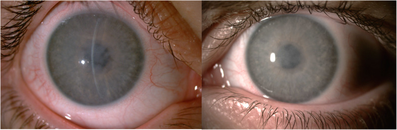
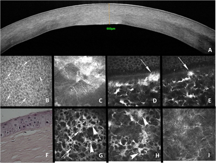
Case presentation: A 16-year-old patient with Hurler's syndrome treated with hematopoietic stem cell transplantation was referred for decreased vision related to advanced keratopathy. The patient was treated with deep anterior lamellar keratoplasty (DALK) in both eyes with uncomplicated outcome. Visual acuity improved from 0.1 (20/200) preoperatively to 0.32 (20/63) and 0.63 (20/32) after transplantation. The corneal endothelial cell density was 2400 cells/mm2 in both eyes 3 years after transplantation. In vivo confocal microscopy (IVCM) and spectral domain optical coherence tomography (SD-OCT) were performed preoperatively. The corneal buttons retrieved during keratoplasty were processed for histology. In SD-OCT scans, corneal opacities appeared as diffuse stromal hyperreflectivity associated with increased corneal thickness. IVCM showed diffuse cytoplasmic granular hyperreflectivity and rounded/ellipsoid aspects of keratocytes, presence of small intracellular vacuoles, and hyperreflective epithelial intercellular spaces. Bowman's layer was thin and irregular. The corneal endothelium was poorly visualized but no endothelial damage was observed. Histology showed irregular orientation and organization of stromal lamellae, with the presence of macrophages whose cytoplasm appeared clear and granular. A perinuclear clear halo was visible within the epithelial basal cells. Bowman's layer featured breaks and irregularities.
Conclusions: The observed corneal multimodal imaging features in mucopolysaccharidosis-related keratopathy were concordant with histology. Compared with standard histology, multimodal imaging allowed additional keratocyte features to be observed. It revealed both morphological and structural changes of all corneal layers but the endothelium. This information is essential for therapeutic management which should include DALK as the first-choice treatment in case of impaired visual acuity.
Keywords: Case report; Deep anterior lamellar keratoplasty; Hurler syndrome; In vivo confocal microscopy; Optical coherence tomography.
Conflict of interest statement
EDC, no financial disclosures. CG, no financial disclosures. NB, no financial disclosures. MP, no financial disclosures. FBB, no financial disclosures. VMB, no financial disclosures.
Figures
References
Publication types
MeSH terms
Grants and funding
LinkOut - more resources
Full Text Sources
Medical
