

Successful laparoscopic surgery combined with selective arterial embolization for bleeding due to jejunal angiodysplasia: a case report
- PMID: 33129311
- PMCID: PMC7603661
- DOI: 10.1186/s12893-020-00924-3
Successful laparoscopic surgery combined with selective arterial embolization for bleeding due to jejunal angiodysplasia: a case report
Abstract
Background: Angiodysplasia of the gastrointestinal tract is a rare vascular pathology that sometimes causes massive hemorrhage. Angiodysplasias are particularly difficult to find in the small intestine for anatomical reasons, often impeding their diagnosis and treatment. Lesion localization is a major challenge in cases of small bowel bleeding requiring surgical intervention.
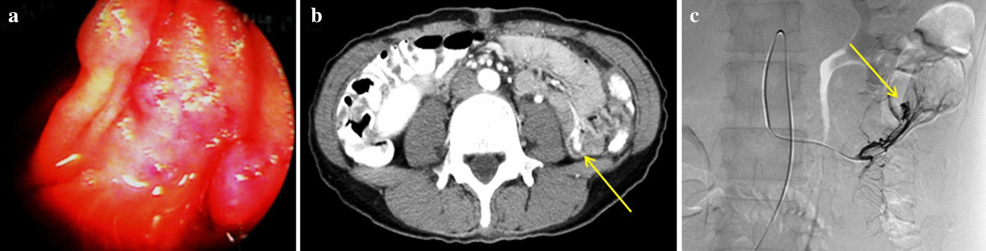
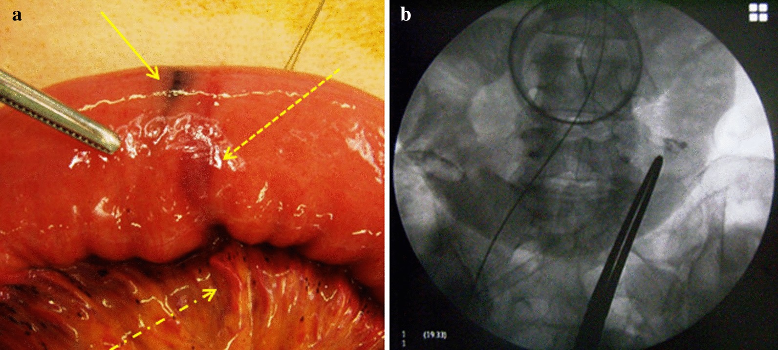
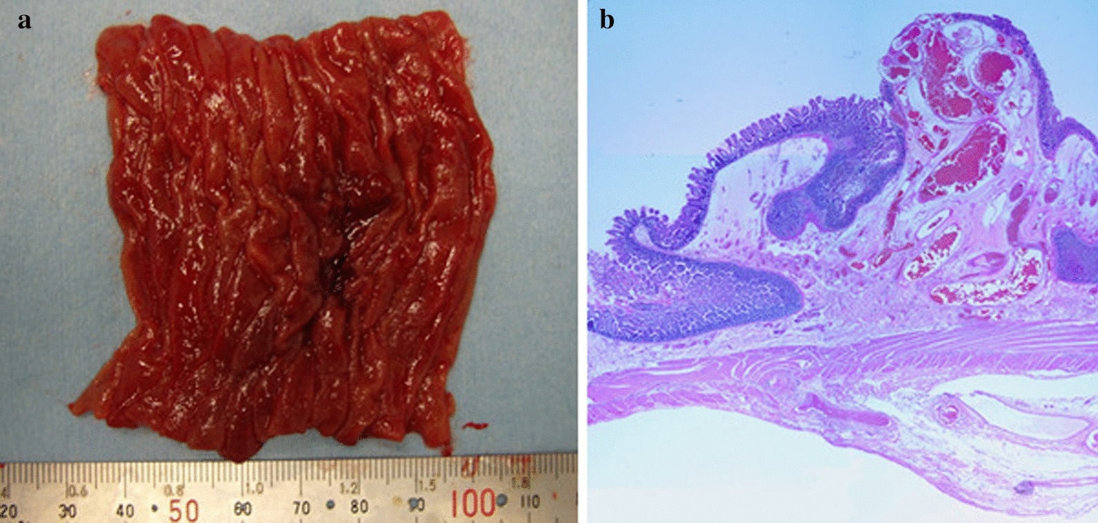
Case presentation: The present case was a 52-year-old woman who was urgently hospitalized with repeated tarry stools. Surgical intervention was chosen after conservative treatment failed to improve her condition. The source of bleeding was suspected to be a vascular lesion discovered in the small intestine during a past double-balloon endoscopy. Abdominal contrast computed tomography revealed a jejunal hemorrhage. We chose selective arterial embolization to stabilize her hemodynamics followed by surgical intervention as her treatment plan. Several embolic and contrast agents (cyanoacrylate, indigo carmine, and Lipiodol) were combined to help identify the location of the lesion during surgery. This multi-pronged approach allowed us to localize the lesion under laparoscopic guidance with high confidence and accuracy, and to excise a 6-cm segment of the small intestine. The lesion was histologically diagnosed as angiodysplasia. No re-bleeding has been observed since the operation.
Conclusion: We report our experience with a case of jejunal angiodysplasia, which was localized with selective arterial embolization using an array of embolic and contrast agents, and then excised laparoscopically. Selective arterial embolization with indigo carmine dye to treat small bowel bleeding preoperatively not only makes the surgery safer by stabilizing the patient's hemodynamics, but is also very useful for localizing the lesion intraoperatively.
Keywords: Indigo carmine; Laparoscopic surgery; Selective arterial embolization; Small bowel bleeding; Small intestinal angiodysplasia.
Conflict of interest statement
The authors declare that they have no competing interests.
Figures
Similar articles
-
[Jejunal phlebectasia as a cause of massive gastrointestinal haemorrhage].Rev Gastroenterol Peru. 2019 Jan-Mar;39(1):84-87. Rev Gastroenterol Peru. 2019. PMID: 31042243 Spanish.
-
[Jejunal angiodysplasia as a cause of obscure gastrointestinal bleeding].Cir Cir. 2008 May-Jun;76(3):261-4. Cir Cir. 2008. PMID: 18647562 Spanish.
-
[Preoperative identification of bleeding site caused by angiodysplasia of the small bowel by means of selective arteriography and application of methylene blue].Vojnosanit Pregl. 2010 Dec;67(12):1029-32. doi: 10.2298/vsp1012029s. Vojnosanit Pregl. 2010. PMID: 21425561 Serbian.
-
Jejunal angiodysplasia confirmed by intravascular injection technique in vitro. Report of a case and review of the literature.J Clin Gastroenterol. 1996 Sep;23(2):139-44. doi: 10.1097/00004836-199609000-00017. J Clin Gastroenterol. 1996. PMID: 8877645 Review.
-
Paediatric angiodysplasia of the jejunum: a case report and review of the literature.Scott Med J. 2012 Nov;57(4):247. doi: 10.1258/smj.2012.012082. Epub 2012 Sep 18. Scott Med J. 2012. PMID: 22989993 Review.
Cited by
-
Jejunal angiodysplasia: surgery can be life-saving - a case report.Ann Med Surg (Lond). 2024 Feb 28;86(4):2204-2207. doi: 10.1097/MS9.0000000000001799. eCollection 2024 Apr. Ann Med Surg (Lond). 2024. PMID: 38576924 Free PMC article.
-
Thermal and Magnetic Dual-Responsive Catheter-Assisted Shape Memory Microrobots for Multistage Vascular Embolization.Research (Wash D C). 2024 Mar 26;7:0339. doi: 10.34133/research.0339. eCollection 2024. Research (Wash D C). 2024. PMID: 38550780 Free PMC article.
-
Jejunal Angiodysplasia as an Unusual Cause of Hemorrhage After Hepaticojejunostomy.Cureus. 2024 Jun 16;16(6):e62502. doi: 10.7759/cureus.62502. eCollection 2024 Jun. Cureus. 2024. PMID: 39022469 Free PMC article.
References
Publication types
MeSH terms
LinkOut - more resources
Full Text Sources
Medical
Miscellaneous
