. 2020 Oct 31;13(1):542.
doi: 10.1186/s13071-020-04300-1.

Identification key to the Anopheles mosquitoes of South America (Diptera: Culicidae). III. Male genitalia

Affiliations

Identification key to the Anopheles mosquitoes of South America (Diptera: Culicidae). III. Male genitalia

Maria Anice Mureb Sallum et al. Parasit Vectors. .

Abstract

Background: Accurate identification of the species of Anopheles Meigen, 1818 requires careful examination of all life stages. However, morphological characters, especially those of the females and fourth-instar larvae, show some degree of polymorphism and overlap among members of species complexes, and sometimes even within progenies. Characters of the male genitalia are structural and allow accurate identification of the majority of species, excluding only those in the Albitarsis Complex. In this key, based on the morphology of the male genitalia, traditionally used important characters are exploited together with additional characters that allow robust identification of male Anopheles mosquitoes in South America.

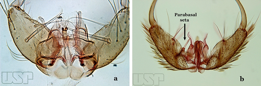
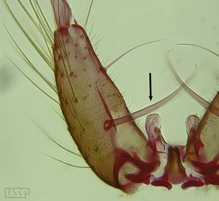
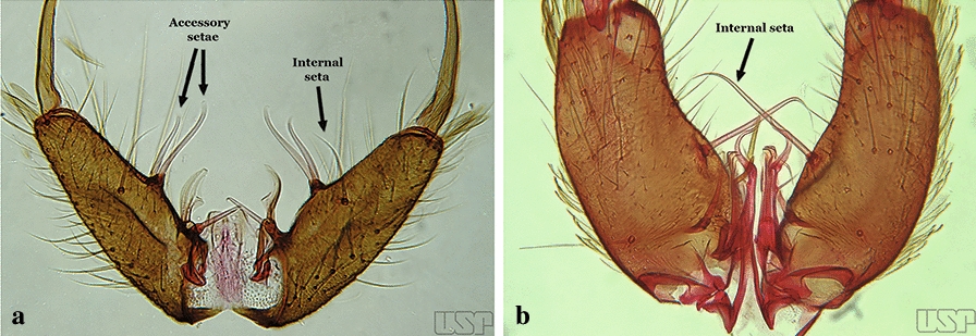
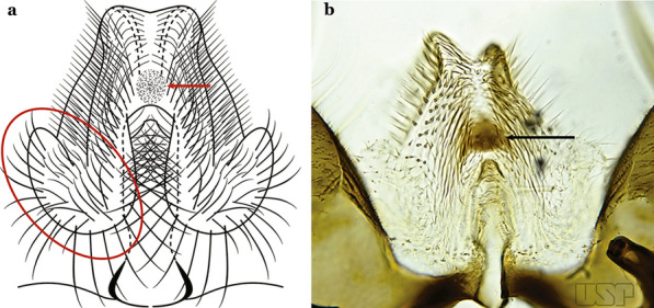
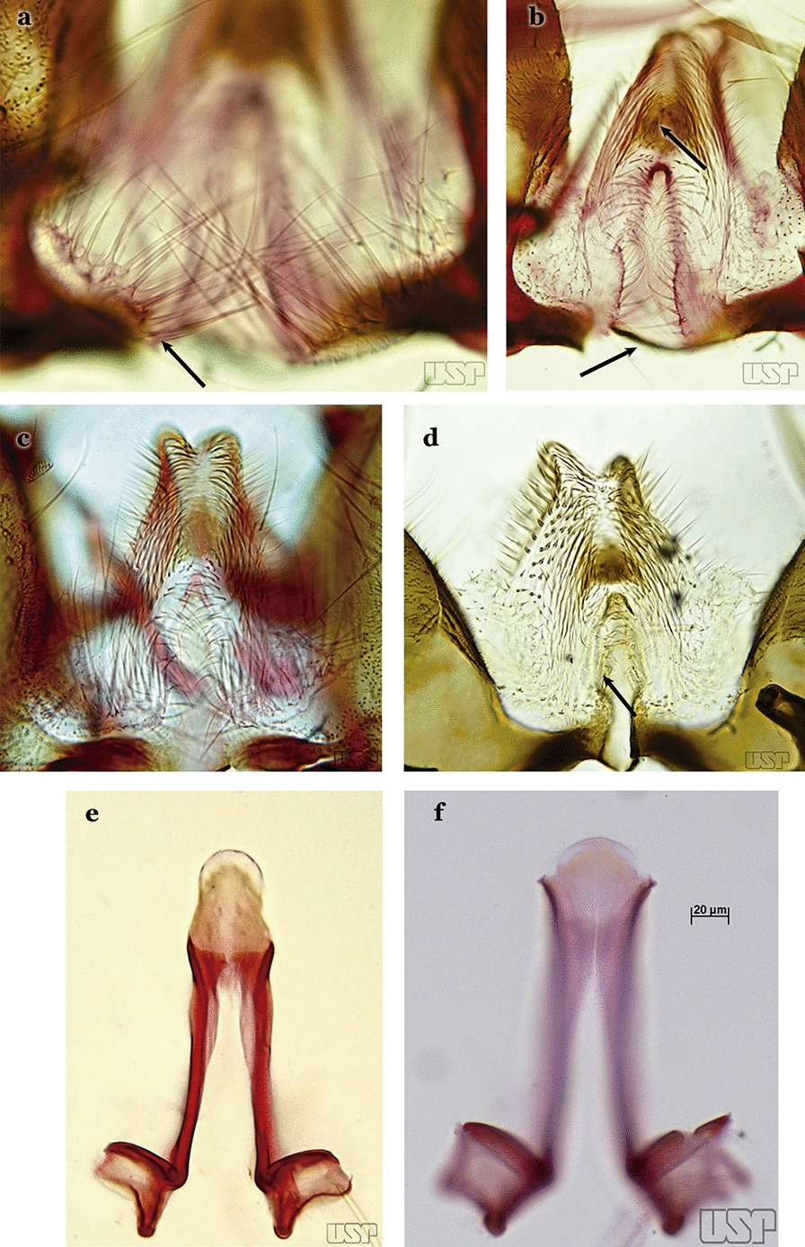
Methods: Morphological characters of the male genitalia of South American species of the genus Anopheles were examined and employed to construct a comprehensive, illustrated identification key. For those species for which specimens were not available, illustrations were based on published illustrations. Photographs of key characters of the genitalia were obtained using a digital Canon Eos T3i attached to a light Diaplan Leitz microscope. The program Helicon Focus was used to build single in-focus images by stacking multiple images of the same structure.

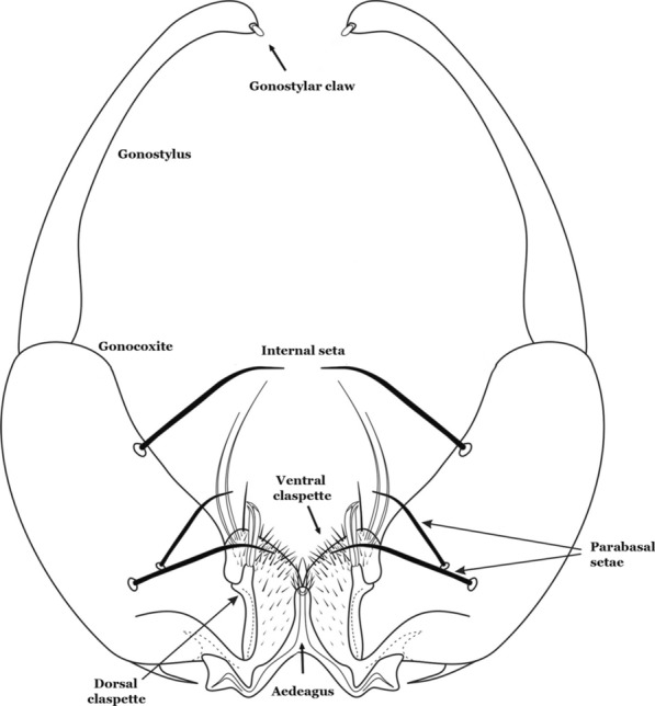
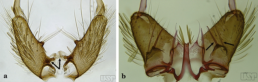
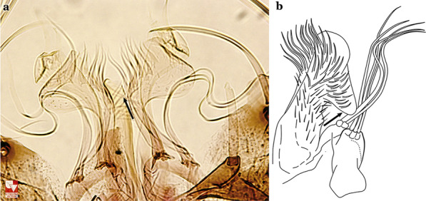
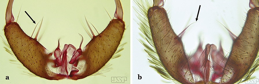
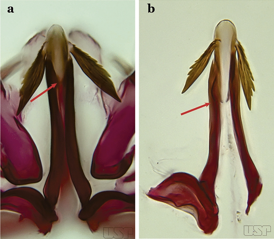
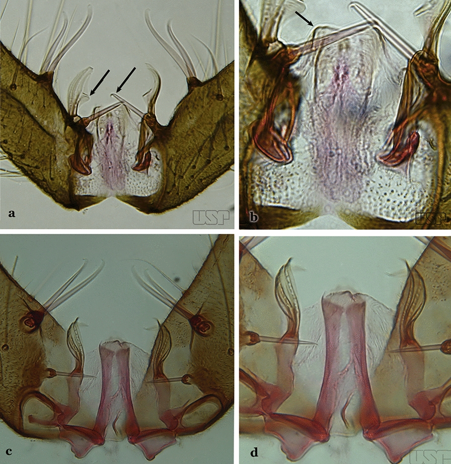
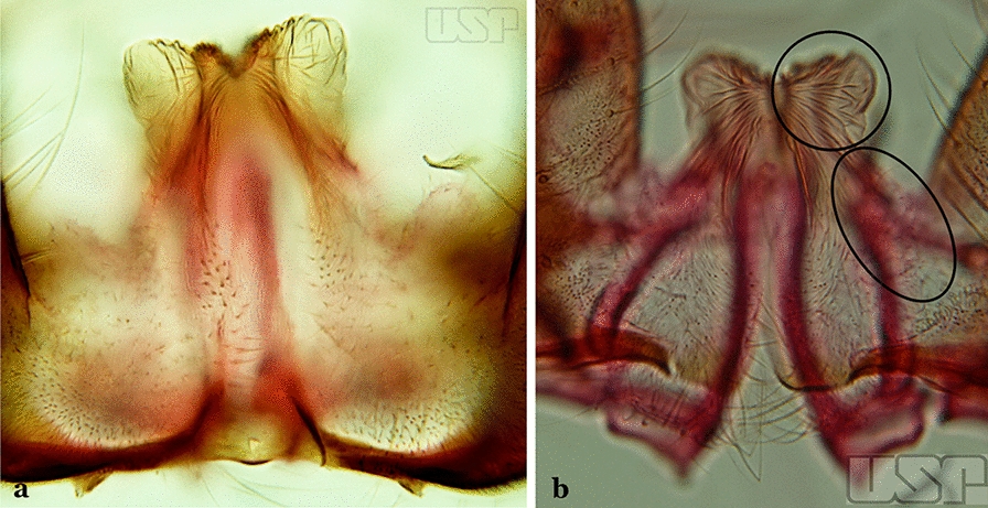
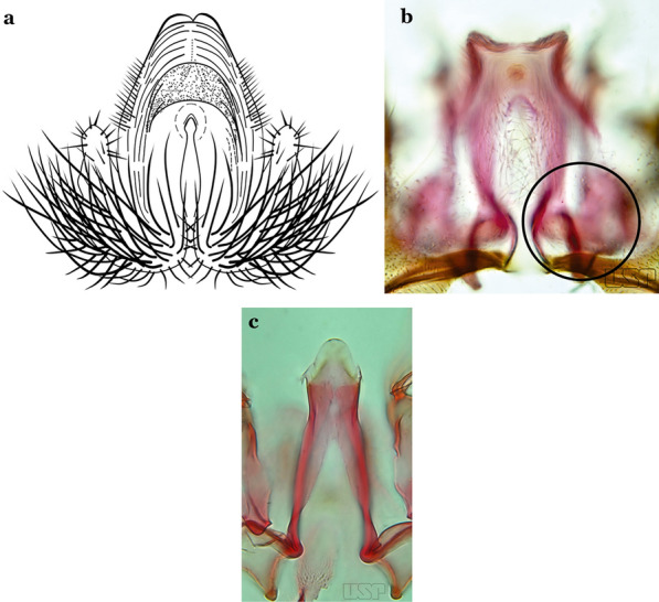
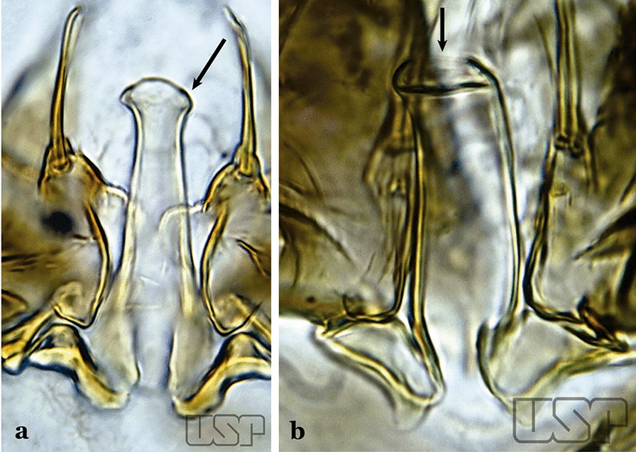
Results: An illustrated key to South American species of Anopheles based on the morphology of the male genitalia is presented, together with a glossary of morphological terms. The male genitalia of type-specimens of previously poorly documented species were also examined and included in the key, e.g. Anopheles (Anopheles) tibiamaculatus (Neiva, 1906) which has a unique quadrangular-shaped aedeagus with an apical opening.

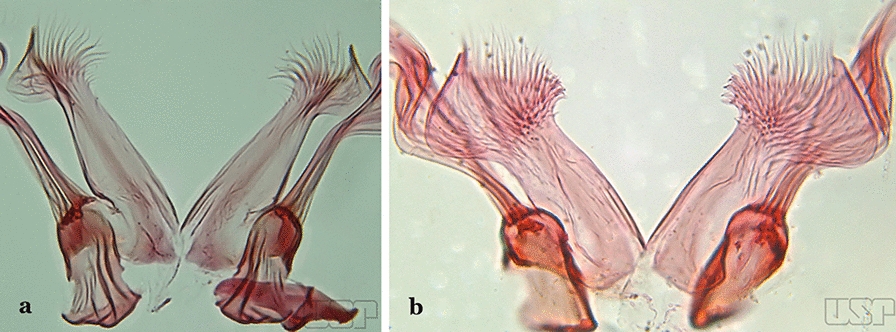
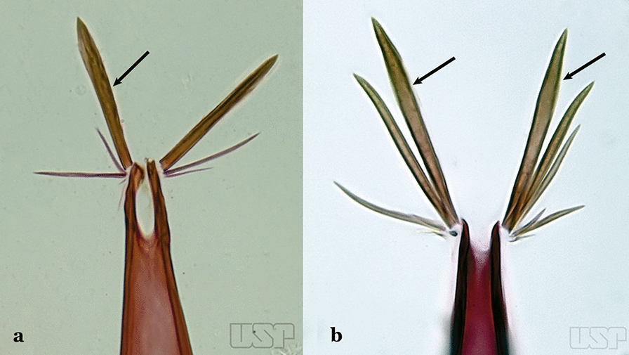
Conclusions: Male genitalia of South American species of Anopheles possess robust characters that can be exploited for accurate species identification. Distortion that can occur during the dissection and mounting process can obstruct accurate identification; this is most evident with inadvertent damage or destruction of unique features and interferes with correctly assigning shapes of the features of the ventral claspette. In some species, the shape, and anatomical details of the aedeagus also need to be examined for species identification. For members of the Myzorhynchella Series, both ventral and dorsal claspettes possess multiple characteristics that are herein used as reliable characters for species identification.

Keywords: Anopheles; Illustrated key; Male genitalia; Morphology; South America.


Conflict of interest statement

The authors declare that they have no competing interests.

Figures

Fig. 1
Fig. 1
Schematic drawing of the male genitalia of An. pseudopunctipennis Theobald, 1901. The proctiger (anal lobe) was removed to permit an unobstructed view of the ventral structures (redrawn after Komp [7])
Fig. 2
Fig. 2
Dorsal view of the male genitalia of An. albimanus Wiedemann. Segment IX and the proctiger (anal lobe) were removed to facilitate the observation of structures that occupy the ventral position
Fig. 3
Fig. 3
Gonocoxites showing the accessory setae, internal seta and parabasal setae of species of the subgenera Nyssorhynchus (a), Anopheles (b), Stethomyia (c). b and c redrawn after Komp [7]
Fig. 4
Fig. 4
Ventral and dorsal lobe claspettes of the male of An. neomaculipalpus Curry, 1931(redrawn after Komp [7])
Fig. 5
Fig. 5
Ventral claspette of An. strodei Root, 1926 (redrawn after Faran & Linthicum [10])
Fig. 6
Fig. 6
Basal portion of dorsal claspette of species of Anopheles (Nyssorhynchus). a An. braziliensis (Chagas, 1907). b An. argyritarsis Robineau-Desvoidy, 1827 (redrawn after Faran & Linthicum [10])
Fig. 7
Fig. 7
Aedeagus of the male genitalia. a Subgenus Anopheles. b Subgenus Nyssorhynchus (redrawn after Komp [7])
Fig. 8
Fig. 8
aAn. thomasi Shannon, 1933. b An. antunesi Galvao & Franco do Amaral, 1940
Fig. 9
Fig. 9
aAn. kompi Edwards, 1930. b An. thomasi
Fig. 10
Fig. 10
An. kompi
Fig. 11
Fig. 11
aAn. thomasi. bAn. nimbus (Theobald, 1902)
Fig. 12
Fig. 12
aAn. acanthotorynus Komp, 1937 (redrawn after Komp [12]). b, c An. nimbus (c drawn from b)
Fig. 13
Fig. 13
aAn. braziliensis. bAn. costai da Fonseca & da Silva Ramos, 1940
Fig. 14
Fig. 14
aAn. darlingi Root, 1926. b An. peryassui Dyar & Knab, 1908
Fig. 15
Fig. 15
a, bAn. homunculus Komp, 1937. c An. darlingi Root, 1926
Fig. 16
Fig. 16
Aedeagus of An. neivai (Lane & Coutinho, 1940) (a) and An. cruzii Dyar & Knab, 1908 (b)
Fig. 17
Fig. 17
a Ventral claspette of An. bambusicolus Komp, 1937. b, c An. homunculus (b redrawn after Zavortink [13])
Fig. 18
Fig. 18
An. homunculus
Fig. 19
Fig. 19
aAn. neivai. bAn. homunculus
Fig. 20
Fig. 20
aAn. pholidotus Zavortink, 1973. b An. lepidotus Zavortink, 1973 (redrawn after Zavortink [13])
Fig. 21
Fig. 21
a, bAn. homunculus (a redrawn after Zavortink [13]). c An. laneanus Correa & Cerqueira, 1944
Fig. 22
Fig. 22
aAn. cruzii. bAn. bellator Dyar & Knab, 1906
Fig. 23
Fig. 23
aAn. bellator. bAn. laneanus
Fig. 24
Fig. 24
aAn. triannulatus. bAn. strodei
Fig. 25
Fig. 25
aAn. triannulatus. bAn. guarani Shannon, 1928
Fig. 26
Fig. 26
aAn. triannulatus. bAn. halophylus
Fig. 27
Fig. 27
aAn. guarani. bAn. albitarsis Lynch Arribálzaga, 1938
Fig. 28
Fig. 28
aAn. parvus (Chagas, 1907). b An. antunesi Galvão & Franco do Amaral, 1940
Fig. 29
Fig. 29
aAn. atacamensis González & Sallum, 2010. b An. darlingi
Fig. 30
Fig. 30
aAn. antunesi. bAn. atacamensis
Fig. 31
Fig. 31
aAn. antunesi. bAn. pristinus Nagaki & Sallum, 2010
Fig. 32
Fig. 32
aAn. pictipennis (Philippi, 1865). b An. lutzii Cruz, 1901
Fig. 33
Fig. 33
aAn. atacamensis. bAn. pictipennis
Fig. 34
Fig. 34
a, bAn. lutzii. c, dAn. guarani Shannon, 1928
Fig. 35
Fig. 35
a, bAn. darlingi. c, d, eAn. lanei Galvão & Franco do Amaral, 1938
Fig. 36
Fig. 36
aAn. lanei. b, cAn. argyritarsis
Fig. 37
Fig. 37
aAn. sawyeri Causey, Deane, Deane & Sampaio, 1943. b An. argyritarsis
Fig. 38
Fig. 38
aAn. albimanus (redrawn after Faran [9]). b An. marajoara Galvão & Damasceno, 1942
Fig. 39
Fig. 39
aAn. albitarsis Lynch Arribálzaga, 1878. b An. marajoara
Fig. 40
Fig. 40
a, bAn. braziliensis. c, dAn. marajoara
Fig. 41
Fig. 41
aAn. strodei. bAn. nuneztovari Gabaldon, 1940
Fig. 42
Fig. 42
a, bAn. benarrochi Gabaldon, Cova-Garcia & Lopez, 1941. c An. rondoni (Neiva & Pinto, 1922). d An. strodei
Fig. 43
Fig. 43
a, bAn. rondoni. c, dAn. strodei
Fig. 44
Fig. 44
aAn. arthuri Unti, 1941. b An. albertoi Unti, 1941
Fig. 45
Fig. 45
aAn. strodei. bAn. albertoi
Fig. 46
Fig. 46
aAn. ininii (redrawn after Faran [9]). b An. goeldii Rozeboom & Gabaldon, 1941
Fig. 47
Fig. 47
aAn. ininii Senevet & Abonnenc, 1938 (redrawn after Faran [9]). b, c An. nuneztovari
Fig. 48
Fig. 48
An. goeldii
Fig. 49
Fig. 49
aAn. rangeli Gabaldon, Cova-Garcia & Lopez, 1940. b An. dunhami Causey, 1945
Fig. 50
Fig. 50
aAn. dunhami. bAn. galvaoi Causey, Deane & Deane, 1943
Fig. 51
Fig. 51
aAn. evansae (Brèthes, 1926). b An. galvaoi
Fig. 52
Fig. 52
aAn. aquasalis Curry, 1932 (redrawn after Faran [9]). b An. konderi Galvão & Damasceno, 1942
Fig. 53
Fig. 53
a, bAn. galvaoi. c, eAn. oswaldoi (Peryassú, 1922). d, f An. konderi
Fig. 54
Fig. 54
aAn. squamifemur Antunes, 1937. b An. pseudotibiamaculatus Galvão & Barretto, 1941
Fig. 55
Fig. 55
aAn. pseudotibiamaculatus. bAn. gilesi (Neiva, 1908)
Fig. 56
Fig. 56
aAn. pseudotibiamaculatus. bAn. gomezdelatorrei Levi-Castillo, 1955
Fig. 57
Fig. 57
aAn. vargasi Gabaldon, Cova-Garcia & Lopez, 1941 (redrawn after Levi-Castillo [15]). b An. gilesi (Neiva, 1908)
Fig. 58
Fig. 58
a, bAn. gilesi. c, dAn. oiketorakras Osorno-Mesa, 1947 redrawn after Lane [16]
Fig. 59
Fig. 59
aAn. tibiamaculatus Neiva, 1906. b An. costai
Fig. 60
Fig. 60
aAn. mediopunctatus (Lutz, 1903). b An. eiseni Coquillett, 1902
Fig. 61
Fig. 61
a, bAn. mediopunctatus. cAn. costai. dAn. forattinii Wilkerson & Sallum, 1999
Fig. 62
Fig. 62
aAn. costai. bAn. forattinii redrawn after Wilkerson & Sallum [17]
Fig. 63
Fig. 63
aAn. mattogrossensis Lutz & Neiva, 191163. b An. peryassui
Fig. 64
Fig. 64
a, bAn. vestitipennis Dyar & Knab, 1906 (redrawn after Komp [7]). c An. mattogrossensis Lutz & Neiva, 1911
Fig. 65
Fig. 65
aAn. mattogrossensis. b, cAn. eiseni geometricus Correa, 1944
Fig. 66
Fig. 66
aAn. peryassui. bAn. calderoni
Fig. 67
Fig. 67
a, bAn. peryassui. c, dAn. minor da Costa Lima, 1929
Fig. 68
Fig. 68
aAn. anchietai Corrêa & Ramalho, 1968. b An. fluminensis Root, 1927
Fig. 69
Fig. 69
aAn. maculipes (Theobald, 1903). b An. anchietai
Fig. 70
Fig. 70
aAn. apicimacula Dyar & Knab, 1906. b An. pseudopunctipennis (both redrawn after Komp [7])
Fig. 71
Fig. 71
aAn. apicimacula. b, cAn. punctimacula Dyar & Knab, 1906 (both redrawn after Komp [7])
Fig. 72
Fig. 72
a, bAn. calderoni Wilkerson, 1991. c, d An. shannoni Davis, 1931 (redrawn after Wilkerson et al. [18])
Fig. 73
Fig. 73
aAn. medialis Harbach, 2018 (redrawn after Wilkerson et al. [18]). b An. fluminensis
Fig. 74
Fig. 74
aAn. medialis. bAn. fluminensis
Fig. 75
Fig. 75
a, bAn. neomaculipalpus (b redrawn from [7], c An. pseudopunctipennis (redrawn after Komp [7])

References

    1. Sallum MAM, Gonzalez Obando R, Carrejo N, Wilkerson RC. Identification keys to the Anopheles mosquitoes of South America (Diptera: Culicidae). I. Introduction. Parasit Vectors. 2020 doi: 10.1186/s13071-020-04298-6. - DOI - PMC - PubMed
    1. Sallum MAM, Gonzalez Obando R, Carrejo N, Wilkerson RC. Identification keys to the Anopheles mosquitoes of South America (Diptera: Culicidae). II. Fourth instar larvae. Parasit Vectors. 2020 doi: 10.1186/s13071-020-04299-5. - DOI - PMC - PubMed
    1. Sallum MAM, Gonzalez Obando R, Carrejo N, Wilkerson RC. Identification keys to the Anopheles mosquitoes of South America (Diptera: Culicidae). IV. Adult females. Parasit Vectors. 2020 doi: 10.1186/s13071-020-04301-0. - DOI - PMC - PubMed
    1. Harbach RE, Knight KL. Taxonomistsʼ glossary of mosquito anatomy. Marlton: Plexus Publishing, Inc.; 1980.
    1. Harbach RE, Knight KL. Corrections and additions to taxonomistsʼ glossary of mosquito anatomy. Mosq Syst. 1981;1981:201–217.

LinkOut - more resources