

Sublethal Supraphysiological Shear Stress Alters Erythrocyte Dynamics in Subsequent Low-Shear Flows
- PMID: 33130119
- PMCID: PMC7732817
- DOI: 10.1016/j.bpj.2020.10.022
Sublethal Supraphysiological Shear Stress Alters Erythrocyte Dynamics in Subsequent Low-Shear Flows
Abstract
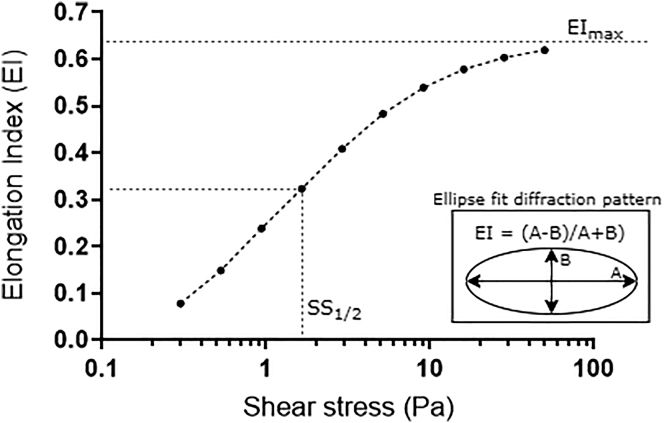
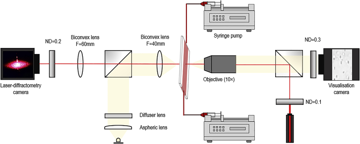
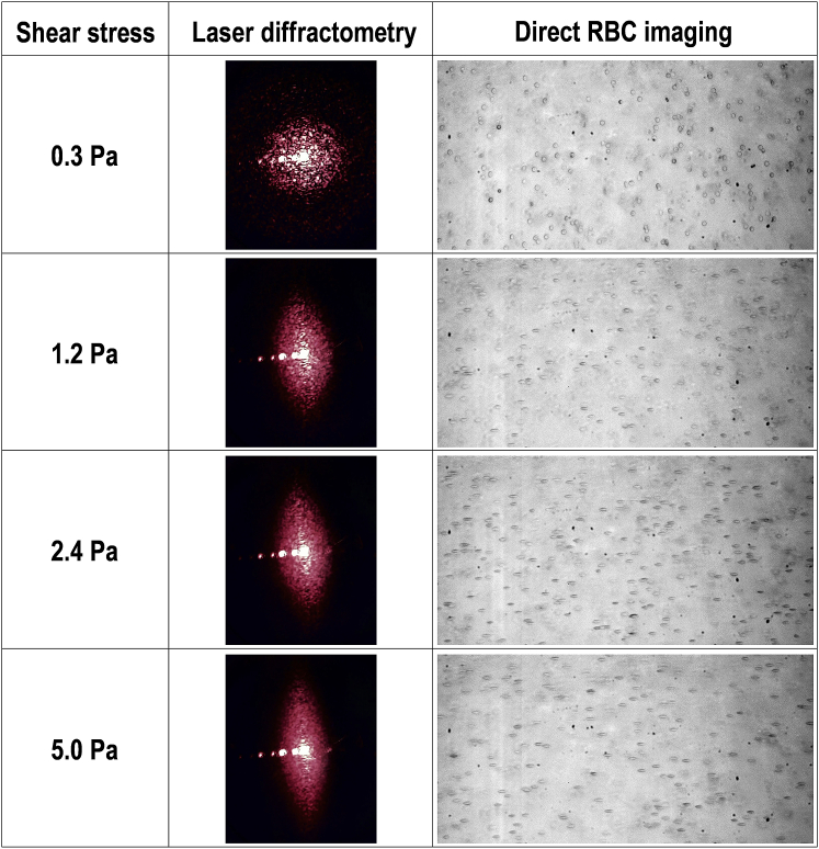
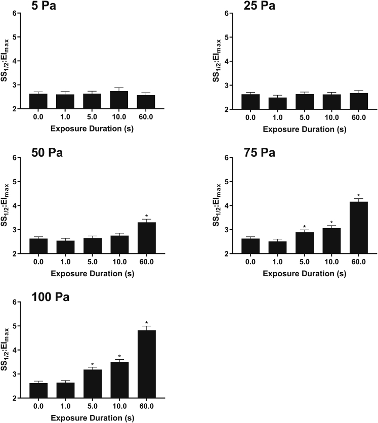
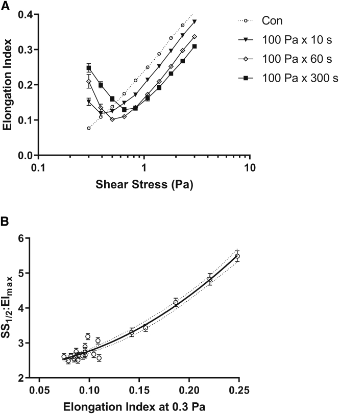
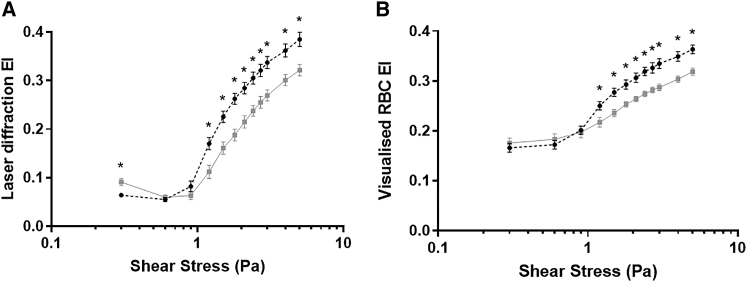
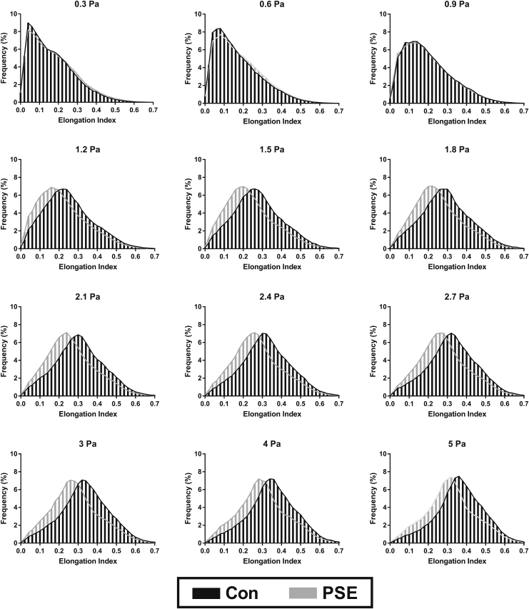
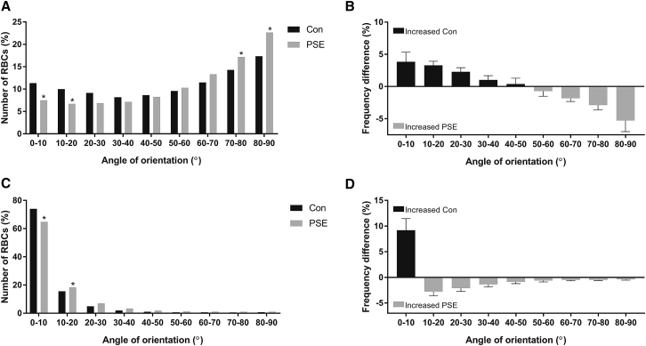
Blood is a non-Newtonian, shear-thinning fluid owing to the physical properties and behaviors of red blood cells (RBCs). Under increased shear flow, pre-existing clusters of cells disaggregate, orientate with flow, and deform. These essential processes enhance fluidity of blood, although accumulating evidence suggests that sublethal blood trauma-induced by supraphysiological shear exposure-paradoxically increases the deformability of RBCs when examined under low-shear conditions, despite obvious decrement of cellular deformation at moderate-to-higher shear stresses. Some propose that rather than actual enhancement of cell mechanics, these observations are "pseudoimprovements" and possibly reflect altered flow and/or cell orientation, leading to methodological artifacts, although direct evidence is lacking. This study thus sought to explore RBC mechanical responses in shear flow using purpose-built laser diffractometry in tandem with direct optical visualization to address this problem. Freshly collected RBCs were exposed to a mechanical stimulus known to drastically alter cell deformability (i.e., prior shear exposure (PSE) to 100 Pa × 300 s). Samples were subsequently transferred to a custom-built slit-flow chamber that combined laser diffractometry with direct cell visualization. Cell suspensions were sheared in a stepwise manner (between 0.3 and 5.0 Pa), with each step being maintained for 15 s. Deformability and cell orientation indices were recorded for small-scatter Fraunhofer diffraction patterns and also visualized RBCs. PSE RBCs had significantly decreased visualized and laser-derived deformability at any given shear stress ≥1 Pa. Novel, to our knowledge, observations demonstrated that PSE RBCs had increased heterogeneity of direct visualized orientation with flow vector at any shear, which may be due to greater vorticity and thus instability in 5-Pa flow compared with unsheared control. These findings indicate that shear exposure and stress-strain history can alter subsequent RBC behavior in physiologically relevant low-shear flows. These findings may yield insight into microvascular disorders in recipients of mechanical circulatory support and individuals with hematological diseases that alter physical properties of blood.
Crown Copyright © 2020. Published by Elsevier Inc. All rights reserved.
Figures
Similar articles
-
Biphasic impairment of erythrocyte deformability in response to repeated, short duration exposures of supraphysiological, subhaemolytic shear stress.Biorheology. 2016 Nov 9;53(3-4):137-149. doi: 10.3233/BIR-15108. Biorheology. 2016. PMID: 27662271
-
Sublethal mechanical shear stress increases the elastic shear modulus of red blood cells but does not change capillary transit velocity.Microcirculation. 2020 Nov;27(8):e12652. doi: 10.1111/micc.12652. Epub 2020 Aug 25. Microcirculation. 2020. PMID: 32738159
-
Oxidative Stress Increases Erythrocyte Sensitivity to Shear-Mediated Damage.Artif Organs. 2018 Feb;42(2):184-192. doi: 10.1111/aor.12997. Epub 2017 Sep 6. Artif Organs. 2018. PMID: 28877350
-
Disturbed blood flow structuring as critical factor of hemorheological disorders in microcirculation.Clin Hemorheol Microcirc. 1998 Dec;19(4):315-25. Clin Hemorheol Microcirc. 1998. PMID: 9972669 Review.
-
Blood rheology and hemodynamics.Semin Thromb Hemost. 2003 Oct;29(5):435-50. doi: 10.1055/s-2003-44551. Semin Thromb Hemost. 2003. PMID: 14631543 Review.
Cited by
-
Shear stress regulation of nanoparticle uptake in vascular endothelial cells.Regen Biomater. 2023 May 2;10:rbad047. doi: 10.1093/rb/rbad047. eCollection 2023. Regen Biomater. 2023. PMID: 37351014 Free PMC article. Review.
-
Mechanotransduction mechanisms in human erythrocytes: Fundamental physiology and clinical significance.Channels (Austin). 2025 Dec;19(1):2556105. doi: 10.1080/19336950.2025.2556105. Epub 2025 Sep 10. Channels (Austin). 2025. PMID: 40929564 Free PMC article. Review.
-
Protocol for inspecting blood cell dynamics with a custom ektacytometer-rheometer apparatus.STAR Protoc. 2022 Apr 7;3(2):101279. doi: 10.1016/j.xpro.2022.101279. eCollection 2022 Jun 17. STAR Protoc. 2022. PMID: 35434656 Free PMC article.
-
Physical Properties of Blood and their Relationship to Clinical Conditions.Front Physiol. 2022 Jul 6;13:906768. doi: 10.3389/fphys.2022.906768. eCollection 2022. Front Physiol. 2022. PMID: 35874542 Free PMC article. Review.
-
Erythrocyte morphological symmetry analysis to detect sublethal trauma in shear flow.Sci Rep. 2021 Dec 7;11(1):23566. doi: 10.1038/s41598-021-02936-2. Sci Rep. 2021. PMID: 34876652 Free PMC article.
References
-
- Baskurt O.K., Meiselman H.J. Blood rheology and hemodynamics. Semin. Thromb. Hemost. 2003;29:435–450. - PubMed
-
- Chien S. Red cell deformability and its relevance to blood flow. Annu. Rev. Physiol. 1987;49:177–192. - PubMed
-
- Schmid-Schöenbein H., Wells R. Fluid drop-like transition of erythrocytes under shear. Science. 1969;165:288–291. - PubMed
-
- Fischer T.M., Stöhr-Lissen M., Schmid-Schönbein H. The red cell as a fluid droplet: tank tread-like motion of the human erythrocyte membrane in shear flow. Science. 1978;202:894–896. - PubMed
MeSH terms
LinkOut - more resources
Full Text Sources
