

Evaluation of a New Model of Care for People with Complications of Diabetic Retinopathy: The EMERALD Study
- PMID: 33130144
- PMCID: PMC7980088
- DOI: 10.1016/j.ophtha.2020.10.030
Evaluation of a New Model of Care for People with Complications of Diabetic Retinopathy: The EMERALD Study
Erratum in
-
Corrigendum.Ophthalmology. 2021 Jul;128(7):1117. doi: 10.1016/j.ophtha.2021.04.013. Ophthalmology. 2021. PMID: 34154726 Free PMC article. No abstract available.
Abstract
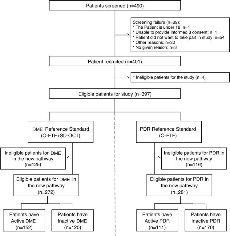
Purpose: The increasing diabetes prevalence and advent of new treatments for its major visual-threatening complications (diabetic macular edema [DME] and proliferative diabetic retinopathy [PDR]), which require frequent life-long follow-up, have increased hospital demands markedly. Subsequent delays in patient's evaluation and treatment are causing sight loss. Strategies to increase capacity are needed urgently. The retinopathy (EMERALD) study tested diagnostic accuracy, acceptability, and costs of a new health care pathway for people with previously treated DME or PDR.
Design: Prospective, multicenter, case-referent, cross-sectional, diagnostic accuracy study undertaken in 13 hospitals in the United Kingdom.
Participants: Adults with type 1 or 2 diabetes previously successfully treated DME or PDR who, at the time of enrollment, had active or inactive disease.
Methods: A new health care pathway entailing multimodal imaging (spectral-domain OCT for DME, and 7-field Early Treatment Diabetic Retinopathy Study [ETDRS] and ultra-widefield [UWF] fundus images for PDR) interpreted by trained nonmedical staff (ophthalmic graders) to detect reactivation of disease was compared with the current standard care (face-to-face examination by ophthalmologists).
Main outcome measures: Primary outcome: sensitivity of the new pathway.
Secondary outcomes: specificity; agreement between pathways; costs; acceptability; proportions requiring subsequent ophthalmologist assessment, unable to undergo imaging, and with inadequate images or indeterminate findings.
Results: The new pathway showed sensitivity of 97% (95% confidence interval [CI], 92%-99%) and specificity of 31% (95% CI, 23%-40%) to detect DME. For PDR, sensitivity and specificity using 7-field ETDRS images (85% [95% CI, 77%-91%] and 48% [95% CI, 41%-56%], respectively) or UWF images (83% [95% CI, 75%-89%] and 54% [95% CI, 46%-61%], respectively) were comparable. For detection of high-risk PDR, sensitivity and specificity were higher when using UWF images (87% [95% CI, 78%-93%] and 49% [95% CI, 42%-56%], respectively, for UWF versus 80% [95% CI, 69-88%] and 40% [95% CI, 34%-47%], respectively, for 7-field ETDRS images). Participants preferred ophthalmologists' assessments; in their absence, they preferred immediate feedback by graders, maintaining periodic ophthalmologist evaluations. When compared with the current standard of care, the new pathway could save £1390 per 100 DME visits and between £461 and £1189 per 100 PDR visits.
Conclusions: The new pathway has acceptable sensitivity and would release resources. Users' suggestions should guide implementation.
Keywords: 7-Field ETDRS images; DME; Diabetes; Early Treatment Diabetic Retinopathy Study; Follow-up; Ophthalmic graders; Ophthalmic photographers; PDR; Pathway; Spectral-domain OCT; Ultra-widefield images.
Copyright © 2020 American Academy of Ophthalmology. All rights reserved.
Figures
Comment in
-
A Holistic Perspective in Caring for People with Diabetic Retinopathy.Ophthalmology. 2021 Apr;128(4):574-575. doi: 10.1016/j.ophtha.2020.12.013. Ophthalmology. 2021. PMID: 33745526 No abstract available.
-
Reply.Ophthalmology. 2021 Sep;128(9):e46-e47. doi: 10.1016/j.ophtha.2021.05.014. Epub 2021 Jun 25. Ophthalmology. 2021. PMID: 34183173 No abstract available.
-
Re: Lois et al.: Evaluation of a new model of care for people with complications of diabetic retinopathy: The EMERALD Study (Ophthalmology. 2021;128:561-573).Ophthalmology. 2021 Sep;128(9):e45-e46. doi: 10.1016/j.ophtha.2021.05.013. Epub 2021 Jun 25. Ophthalmology. 2021. PMID: 34183174 No abstract available.
References
-
- Stitt A.W., Curtis T.M., Chen M. The progress in understanding and treatment of diabetic retinopathy. Prog Retin Eye Res. 2016;51:156–186. - PubMed
-
- Pascolini D., Mariotti S.P. Global estimates of visual impairment: 2010. Br J Ophthalmol. 2012;96:614–618. - PubMed
-
- World Health Organization Blindness and vision impairment. https://www.who.int/news-room/fact-sheets/detail/blindness-and-visual-im... October 8, 2020 Accessed 29.06.20.
-
- Early Treatment Diabetic Retinopathy Study Research Group Photocoagulation for diabetic macular edema. Early Treatment Diabetic Retinopathy Study report number 1. Arch Ophthalmol. 1985;103:1796–1806. - PubMed
Publication types
MeSH terms
Grants and funding
LinkOut - more resources
Full Text Sources
Other Literature Sources
Medical
