Review
. 2021 Feb:151:56-71.
doi: 10.1016/j.yjmcc.2020.10.012. Epub 2020 Oct 29.

Comprehensive evaluation of electrophysiological and 3D structural features of human atrial myocardium with insights on atrial fibrillation maintenance mechanisms

Affiliations
Review

Comprehensive evaluation of electrophysiological and 3D structural features of human atrial myocardium with insights on atrial fibrillation maintenance mechanisms

Aleksei V Mikhailov et al. J Mol Cell Cardiol. 2021 Feb.

Abstract

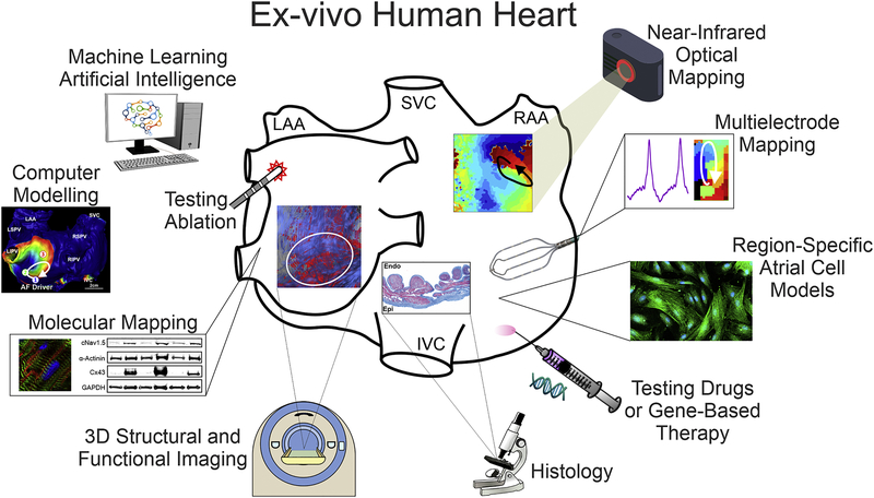
Atrial fibrillation (AF) occurrence and maintenance is associated with progressive remodeling of electrophysiological (repolarization and conduction) and 3D structural (fibrosis, fiber orientations, and wall thickness) features of the human atria. Significant diversity in AF etiology leads to heterogeneous arrhythmogenic electrophysiological and structural substrates within the 3D structure of the human atria. Since current clinical methods have yet to fully resolve the patient-specific arrhythmogenic substrates, mechanism-based AF treatments remain underdeveloped. Here, we review current knowledge from in-vivo, ex-vivo, and in-vitro human heart studies, and discuss how these studies may provide new insights on the synergy of atrial electrophysiological and 3D structural features in AF maintenance. In-vitro studies on surgically acquired human atrial samples provide a great opportunity to study a wide spectrum of AF pathology, including functional changes in single-cell action potentials, ion channels, and gene/protein expression. However, limited size of the samples prevents evaluation of heterogeneous AF substrates and reentrant mechanisms. In contrast, coronary-perfused ex-vivo human hearts can be studied with state-of-the-art functional and structural technologies, such as high-resolution near-infrared optical mapping and contrast-enhanced MRI. These imaging modalities can resolve atrial arrhythmogenic substrates and their role in reentrant mechanisms maintaining AF and validate clinical approaches. Nonetheless, longitudinal studies are not feasible in explanted human hearts. As no approach is perfect, we suggest that combining the strengths of direct human atrial studies with high fidelity approaches available in the laboratory and in realistic patient-specific computer models would elucidate deeper knowledge of AF mechanisms. We propose that a comprehensive translational pipeline from ex-vivo human heart studies to longitudinal clinically relevant AF animal studies and finally to clinical trials is necessary to identify patient-specific arrhythmogenic substrates and develop novel AF treatments.

Keywords: Atrial fibrillation; Electrophysiology; Ex-vivo human heart; Fibrosis; Near-infrared optical mapping.


Figures

Figure 1.
Figure 1.. Advantages of Integrated Explanted Human Heart Approach
Ex-vivo human heart provides multiple advantages over in-vitro single cell and in-vivo studies of the human heart and allows for integration of molecular mapping, histology validated high-resolution 3D structural imaging, standard clinical electrode mapping, and near-infrared optical mapping with subsequent utilization of machine learning and computer modeling. In addition, cellular studies, drug testing, and gene-based therapy, as well as clinical ablation techniques can be utilized to define AF mechanisms and develop patient-specific therapy. Abbreviations are as follows: LAA/RAA – left and right atrial appendages; IVC/SVC – inferior and superior vena cava; Endo/Epi – endocardial, epicardial.
Figure 2.
Figure 2.. Human atrial electrophysiological features evaluated in-vivo, in-vitro and ex-vivo by different methods
A. Action potential recordings using in-vivo, in-vitro single cell, and ex-vivo human heart methods. Adapted from Narayan et al. 2008 [119]; Dobrev et al. 2001 [120]; Zhao et al 2017 [113]. B. In-vivo and ex-vivo CV measurements during pacing. Right: Ex-vivo optical mapping revealed endocardial (Endo) vs. epicardial (Epi) conduction blocks during fast atrial pacing. Adapted from Voskoboinik et al. 2019 [49]; Zhao et al 2017 [113]; Hansen et al. 2018 [77]. C. Left: In-vivo and ex-vivo APD restitution curves. Right: Panoramic ex-vivo APD map of the intact atria. Adapted from Franz et al. 1997 [117]; Zhao et al 2017 [113]. D. In-vivo and ex-vivo dominant frequency (DF) mapping during AF. Left: In-vivo endocardial and epicardial DF maps from electrode recordings in persistent AF patients. Right: Ex-vivo DF map during sustained AF showing stable regions of reentry in areas with high DF in human RA and LA. Adapted from Sanders et al. 2005 [129]; Schuessler et al. 2006 [128]; Zhao et al. 2015 [75]; Li et al., 2016 [57]. Abbreviations are as follows: AF – atrial fibrillation; APD – action potential duration; CV – conduction velocity; DF – dominant frequency; EG - electrogram; ECG – electrocardiogram; IAS – interatrial septum; IVC/SVC – inferior and superior vena cava; LA/RA – left and right atria; LIPV/LSPV/RIPV/RSPV – left, right, superior, inferior pulmonary veins; LRA – left right atria; MAP – monophasic action potential; NIOM – near-infrared optical imaging; OAP – optical action potential; PLA – posterior left atrium; SR – sinus rhythm.
Figure 3.
Figure 3.. Structural substrates of human AF
A. Evaluation of atrial fibrosis by histology and LGE-MRI. Left to right: Fibrosis is upregulated in association with AF in comparison to patients with no history of AF by histology. In-vivo studies showed that the extent of atrial fibrosis was a significant predictor of ablation failure, and likelihood of AF reentrant mechanism increases in areas of high enhancement. Adapted from Platonov et al.2011 [152]; Marrouche et al. 2017 [10, 172]; Cochet et al. 2018 [12]. B. Arrhythmogenic 3D structural substrates in AF driver maintenance. From left to right: Reentrant AF driver identified optically overlapped with 3D CE-MRI anatomy. Bi-atrial wall thickness variations, transmural 3D fibrosis distribution (sub-epi (blue), mid-wall (green), and sub-endo (red)) as well as 3D myofiber tracts created arrhythmogenic hubs for reentrant AF drivers. Adapted from Zhao et al 2017 [113]. C. Histological validation improves ex-vivo and in-vivo fibrosis MRI analysis. Ex-vivo 3D CE-MRI reconstruction shows fibrosis in red and myofibers in blue. In-vivo 3D CE-MRI shows fibrosis islands in possible driver regions. Adapted from Hansen et al. 2015, 2017 [8, 77]; Csepe et al. 2017 [180]. Abbreviations AF – atrial fibrillation; LGE-MRI – late gadolinium enhanced magnetic resonance imaging; IVC/SVC –inferior and superior vena cava; LA/RA – left and right atria; LAA –left atrial appendage; LIPV/LSPV/RIPV/RSPV – left, right, superior, inferior pulmonary veins; CE-MRI – contrast enhanced magnetic resonance imaging; PLA –posterior left atrium
Figure 4.
Figure 4.. Human AF mapping and evolution of AF mechanisms
A. In-vivo epicardial and endocardial mapping visualized different mechanisms of AF maintenance. Adapted from Cox et al.,1991 [165]; Alessie et al., 2014 [170]; Narayan et al., 2013 [169]. B. Dual sided ex-vivo NIOM integrated with clinical multielectrode mapping (MEM) to validate clinical mapping methods and reveal AF driver mechanism. NIOM resolves intramural reentry, which is seen as multiple breakthroughs by clinical epicardial MEM. AF driver denoted by white arrow on Endo view, while star denotes breakthrough visualization on Epi view. Adapted from Hansen et al., 2015, 2018 [77, 83]; Li et al. 2016 [57]. C. Left, progressive electrophysiological and structural remodeling governs AF mechanism. Right, graph showing that the number of AF drivers may depend on the severity of fibrotic remodeling. Abbreviations: Epi – epicardial, Endo – endocardial; AF –atrial fibrillation; FIRM – focal impulse and rotor mapping; MEM – multielectrode mapping; MV – mitral valve; TV – tricuspid valve; LAA/RAA – left and right atrial appendages; NIOM – near-infrared optical mapping; pAF/perAF/LS-perAF – paroxysmal/ persistent AF/long-standing persistent AF; IVC/SVC – inferior and superior vena cava.

Similar articles

Cited by

References

    1. Nishida K, Nattel S, Atrial fibrillation compendium: historical context and detailed translational perspective on an important clinical problem, Circ. Res 114(9) (2014) 1447–1452. - PubMed
    1. Wyse DG, Van Gelder IC, Ellinor PT, Go AS, Kalman JM, Narayan SM, et al., Lone Atrial Fibrillation: Does it Exist?, J. Am. Coll. Cardiol 63(17) (2014) 1715–1723. - PMC - PubMed
    1. Pedersen OD, Abildstrom SZ, Ottesen MM, Rask-Madsen C, Bagger H, Kober L, et al., Increased risk of sudden and non-sudden cardiovascular death in patients with atrial fibrillation/flutter following acute myocardial infarction, Eur Heart J 27(3) (2006) 290–5. - PubMed
    1. Ling LH, Kistler PM, Kalman JM, Schilling RJ, Hunter RJ, Comorbidity of atrial fibrillation and heart failure, Nat Rev Cardiol 13(3) (2016) 131–47. - PubMed
    1. Woods CE, Olgin J, Atrial fibrillation therapy now and in the future: drugs, biologicals, and ablation, Circ. Res 114(9) (2014) 1532–1546. - PMC - PubMed

Publication types

MeSH terms