. 2021 Jan 1:119:155-168.
doi: 10.1016/j.actbio.2020.10.042. Epub 2020 Oct 29.

Xenogeneic-free generation of vascular smooth muscle cells from human induced pluripotent stem cells for vascular tissue engineering

Affiliations

Xenogeneic-free generation of vascular smooth muscle cells from human induced pluripotent stem cells for vascular tissue engineering

Jiesi Luo et al. Acta Biomater. .

Abstract

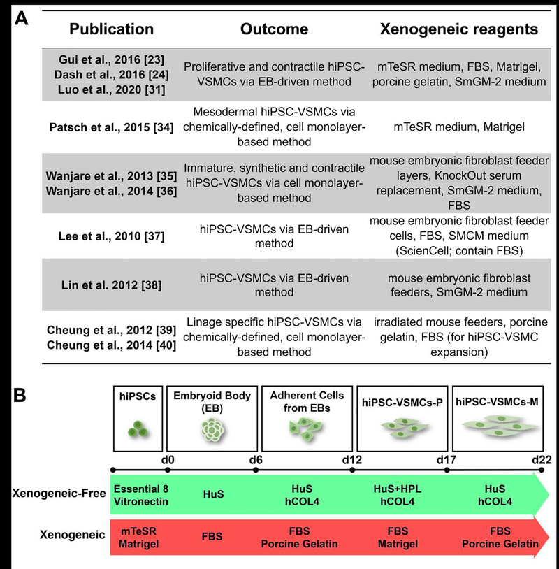
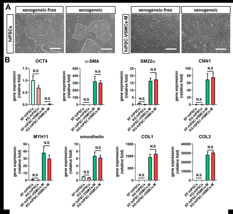
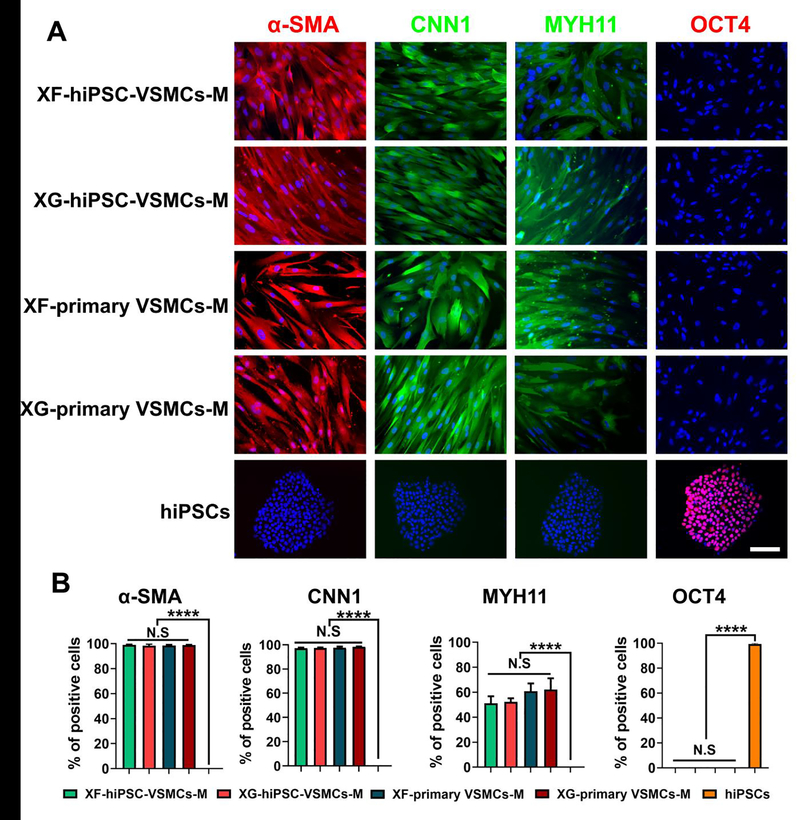
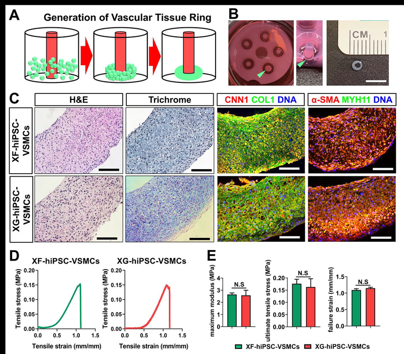
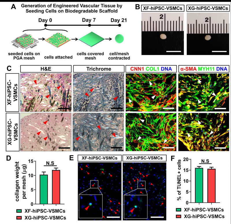
Development of mechanically advanced tissue-engineered vascular grafts (TEVGs) from human induced pluripotent stem cell (hiPSC)-derived vascular smooth muscle cells (hiPSC-VSMCs) offers an innovative approach to replace or bypass diseased blood vessels. To move current hiPSC-TEVGs toward clinical application, it is essential to obtain hiPSC-VSMC-derived tissues under xenogeneic-free conditions, meaning without the use of any animal-derived reagents. Many approaches in VSMC differentiation of hiPSCs have been reported, although a xenogeneic-free method for generating hiPSC-VSMCs suitable for vascular tissue engineering has yet to be established. Based on our previously established standard method of xenogeneic VSMC differentiation, we have replaced all animal-derived reagents with functional counterparts of human origin and successfully derived functional xenogeneic-free hiPSC-VSMCs (XF-hiPSC-VSMCs). Next, our group developed tissue rings via cellular self-assembly from XF-hiPSC-VSMCs, which exhibited comparable mechanical strength to those developed from xenogeneic hiPSC-VSMCs. Moreover, by seeding XF-hiPSC-VSMCs onto biodegradable polyglycolic acid (PGA) scaffolds, we generated engineered vascular tissues presenting effective collagen deposition which were suitable for implantation into an immunodeficient mice model. In conclusion, our xenogeneic-free conditions for generating hiPSC-VSMCs produce cells with the comparable capacity for vascular tissue engineering as standard xenogeneic protocols, thereby moving the hiPSC-TEVG technology one step closer to safe and efficacious clinical translation.

Keywords: Human induced pluripotent stem cells; vascular smooth muscle cells; vascular tissue engineering; xenogeneic-free.


Conflict of interest statement

Declaration of Competing Interest The authors declare that they have no known competing financial interests or personal relationships that could have appeared to influence the work reported in this paper.

Figures

Figure 1.
Figure 1.. Strategy of deriving hiPSC-VSMCs under xenogeneic-free conditions.
(A) Representative studies of currently established methods for deriving vascular smooth muscle cells from human induced pluripotent stem cells. (B) Schematic illustration of VSMC differentiation of hiPSCs under xenogeneic-free or xenogeneic conditions. Note that the essential reagents applied in current xenogeneic-free and previous xenogeneic methods are listed. See the Materials and Methods section and Table S1 for details of the methods. HuS, human serum; HPL, human platelet lysate; hCOL4, human collagen type 4; FBS, fetal bovine serum.
Figure 2.
Figure 2.. Generation of hiPSC-VSMCs under xenogeneic-free conditions.
Results suggested that the hiPSC-VSMCs could be efficiently generated under xenogeneic-free conditions with comparable morphology and gene expression profiles to those under xenogeneic conditions. (A) Representative images of hiPSCs and mature hiPSC-VSMCs under xenogeneic-free or xenogeneic conditions. Scale bar: 200 μm. (B) qRT-PCR analysis of relative mRNA transcript amounts of pluripotency marker (OCT4) and VSMC contractile (α-SMA, SM22α, CNN1, MYH11 and smoothelin) and ECM (COL1 and COL3) markers in xenogeneic-free or xenogeneic hiPSCs (XF-hiPSCs and XG-hiPSCs) and mature hiPSC-VSMCs (XF-hiPSC-VSMCs-M and XG-hiPSC-VSMCs-M). Values in the y-axis represent fold changes relative to human GAPDH expression. Gene expression in each group was normalized to that of in XF-hiPSCs (One-way ANOVA with Tukey’s multiple comparisons test; Mean values and S.E.M indicated by the error bars are shown; n=3; N.S: not significant).
Figure 3.
Figure 3.. Marker expression of XF-hiPSC-VSMCs.
Results suggested that XF-hiPSC-VSMCs presented comparable VSMC marker expression profiles to those of XG-hiPSC-VSMCs and human primary VSMCs. (A) Immunostaining of VSMC (α-SMA, CNN1, and MYH11) and pluripotency (OCT4) markers in XF-hiPSC-VSMCs-M, XG-hiPSC-VSMCs-M, XF-primary VSMCs-M and XG-primary VSMCs-M in maturation stages, and undifferentiated xenogeneic-free hiPSCs. DNA (nuclear) was counterstained by DAPI. Scale bar: 200 μm. (B) The percentage of cells (XF-hiPSC-VSMCs-M, XG-hiPSC-VSMCs-M, XF-primary VSMCs-M, XG-primary VSMCs-M and xenogeneic-free hiPSCs) positive for VSMC (α-SMA, CNN1, and MYH11) and pluripotency (OCT4) markers from immunostaining (One-way ANOVA with Tukey’s multiple comparisons test; Mean values and S.E.M indicated by error bars are shown; n=3; ****: p<0.0001; N.S: not significant).
Figure 4.
Figure 4.. Contractility analysis of XF-hiPSC-VSMCs.
Results suggested that XF-hiPSC-VSMCs displayed comparable contractililty to those of XG-hiPSC-VSMCs and human primary VSMCs. (A) Contractility of XF-hiPSC-VSMCs-M, XG-hiPSC-VSMCs-M, XF-primary VSMCs-M and XG-primary VSMCs-M in maturation stages in response to 1 mM carbachol (before and after 20 minutes incubation). Representative cells were outlined by the blue and red lines to indicate the surface areas before and after carbachol treatment, respectively. Scale bar: 200 μm. (B) Quantification of reduced cell area of xenogeneic-free or xenogeneic hiPSC-VSMCs-M and human primary VSMCs-M in response to 1 mM carbachol or vehicle control (PBS) (One-way ANOVA with Tukey’s multiple comparisons test; Mean values and S.E.M indicated by the error bars are shown; n=3; ***: p < 0.001; ****: p < 0.0001; N.S: not significant).
Figure 5.
Figure 5.. Determining xenogeneic-free collagen promoting medium.
Results suggested that xenogeneic-free collagen promoting medium could lead to robust VSMC marker expression, collagen synthesis, and proliferation of XF-hiPSC-VSMCs. (A) Immunostaining of VSMC contractile (α-SMA and CNN1) and ECM (COL1) markers in XF-hiPSC-VSMCs-P cultured in xenogeneic-free VSMC growth medium, priming medium (DMEM supplemented with 5% human serum and 5% human platelet lysate) or collagen promoting medium for 6 days, and XG-hiPSC-VSMCs-P cultured in xenogeneic collagen promoting medium for 6 days (see Table S1 for detailed media components). DNA (nuclear) was counterstained by DAPI. Scale bar: 200 μm. (B) qRT-PCR analysis of relative mRNA transcript amounts of VSMC contractile (α-SMA and CNN1) and extracellular matrix (COL1) genes in XF-hiPSC-VSMCs cultured in xenogeneic-free VSMC growth medium, priming medium (DMEM supplemented with 5% human serum and 5% human platelet lysate) or collagen promoting medium, and XG-hiPSC-VSMCs-P cultured in xenogeneic collagen promoting medium. Values in the y-axis represent fold changes relative to human GAPDH expression. Gene expression in each group was normalized to that of in XF-hiPSC-VSMCs-P cultured in xenogeneic-free VSMC growth medium (One-way ANOVA with Tukey’s multiple comparisons test; Mean values and S.E.M indicated by the error bars are shown; n=3; *: p < 0.05; **: p < 0.01; ***: p < 0.001; N.S: not significant). (C) Collagen weight per XF- or XG-hiPSC-VSMCs-P cultured in variant media via a hydroxyproline assay (One-way ANOVA with Tukey’s multiple comparisons test; Mean values and S.E.M indicated by the error bars are shown; n=3; ****: p < 0.0001; N.S: not significant). (D) Proliferation rates of XF- or XG-hiPSC-VSMCs-P cultured in various media for 6 days. Paired two-way ANOVA showed that there was no interaction between the time for cell counting and cell/culture conditions. The cell number significantly increased along with the increase of culturing time, while there was no significant difference between the groups of cell/culture conditions within each day (Mean values and S.E.M indicated by the error bars are shown; N.S: not significant).
Figure 6.
Figure 6.. Fabrication of scaffold-free vascular tissue rings using XF-hiPSC-VSMCs.
Results suggested that XF-hiPSC-VSMCs could be used to develop scaffold-free vascular tissue rings with comparable histological and mechanical properties to those from XG-hiPSC-VSMCs. (A) Illustrative scheme of the method used to establish vascular tissue rings from XF-hiPSC-VSMCs-P. Scale bar: 5 mm. (B) Morphology of engineered tissue rings from XF-hiPSC-VSMCs-P. The tissue ring in a well of the agarose mold from above (left image), at a 45-degree angle (middle image) and the size of the ring were shown (right image). Rings are indicated by the light green arrows. (C) H&E staining, Masson’s trichrome staining and immunofluorescent staining (CNN1, COL1, α-SMA and MYH11) of the engineered vascular tissue rings made from XF- or XG-hiPSC-VSMCs-P under xenogeneic-free or xenogeneic conditions, respectively. DNA (nuclear) was counterstained by DAPI in immunostaining. Scale bar: 100 mm. (D) Representative stress-strain plots of engineered vascular tissue rings made from XF- or XG-hiPSC-VSMCs-P under xenogeneic-free or xenogeneic conditions, respectively. (E) Mechanical parameters (maximum modulus, ultimate tensile stress, and failure strain) were compared between the engineered vascular tissue rings made from XF- or XG-hiPSC-VSMCs-P under xenogeneic-free or xenogeneic conditions, respectively (Two-tailed paired Student’s T-test; Mean values and S.E.M indicated by the error bars are shown; N.S: not significant).
Figure 7.
Figure 7.. Generation of engineered tissues from culturing XF-hiPSC-VSMCs on biodegradable polyglycolic acid scaffolds.
Results suggested that XF-hiPSC-VSMCs could be cultured on biodegradable PGA scaffold to develop engineered vascular tissues with comparable collagen depsotion and histological properties to those from XG-hiPSC-VSMCs. (A) Schematic illustration for developing tissue patches from XF-hiPSC-VSMCs-P grown on PGA scaffolds. Scale bar: 5 mm. (B) Morphology of the engineered tissues developed from XF- or XG-hiPSC-VSMCs-P grown on PGA scaffolds (day 21). (C) H&E staining, Masson’s trichrome staining and immunofluorescent staining (CNN1, COL1, α-SMA and MYH11) of the engineered tissues made from XF- or XG-hiPSC-VSMCs-P under xenogeneic-free or xenogeneic conditions, respectively. DNA (nuclear) was counterstained by DAPI in immunostaining. Red and white arrow heads indicate PGA remnants. Scale bar: 100 mm. (D) Collagen weight per mesh of engineered tissue made from XF- or XG-hiPSC-VSMCs-P under xenogeneic-free or xenogeneic conditions, respectively, via hydroxyproline assay (Two-tailed paired Student’s T-test; Mean values and S.E.M indicated by the error bars are shown; N.S: not significant). (E) TUNEL staining of the engineered tissues from XF- or XG-hiPSC-VSMCs-P under xenogeneic-free or xenogeneic conditions, respectively. DNA (nuclear) was counterstained by DAPI. Red arrow heads indicate the TUNEL-positive apoptotic cells. Scale bar: 100 μm. (F) Quantification of percentage of TUNEL-positive cells in engineered tissues from XF- or XG-hiPSC-VSMCs-P under xenogeneic-free or xenogeneic conditions, respectively (Two-tailed paired Student’s T-test; Mean values and S.E.M indicated by the error bars are shown; N.S: not significant).
Figure 8.
Figure 8.. Engraftment of engineered tissues from culturing XF-hiPSC-VSMCs on PGA scaffolds.
Results suggested that engineered vascular tissues developed using XF-hiPSC-VSMCs presented similar histological properties to those from XG-hiPSC-VSMCs after subcutaneous implantation into immune-deficient mouse model. (A) Schematic illustration for subcutaneously engrafting engineered tissue from XF-hiPSC-VSMCs-P grown on PGA scaffolds. Scale bar: 5 mm. (B) Morphology of the explanted engineered tissues on day 14 post-engraftment. (C) H&E staining, Masson’s trichrome staining and immunofluorescent staining (CNN1, human leukocyte antigen-A [HLA-A], α-SMA and MYH11) of the explanted engineered tissues made from XF- or XG-hiPSC-VSMCs-P under xenogeneic-free or xenogeneic conditions, respectively, or blank control PGA meshes without seeded cells. DNA (nuclear) was counterstained by DAPI in immunostaining. Red arrow heads indicate PGA remnants. White lines indicate the border between the engrafted and host tissues. Scale bar: 100 μm.

Similar articles

Cited by

References

    1. Feliciano DV, Pitfalls in the management of peripheral vascular injuries, Trauma surgery & acute care open 2(1) (2017) e000110. - PMC - PubMed
    1. Feliciano DV, Moore EE, West MA, Moore FA, Davis JW, Cocanour CS, Scalea TM, McIntyre RC Jr., Western Trauma Association critical decisions in trauma: evaluation and management of peripheral vascular injury, part II, The journal of trauma and acute care surgery 75(3) (2013) 391–7. - PubMed
    1. Pashneh-Tala S, MacNeil S, Claeyssens F, The Tissue-Engineered Vascular Graft-Past, Present, and Future, Tissue Eng Part B Rev 22(1) (2016) 68–100. - PMC - PubMed
    1. Schild AF, Perez E, Gillaspie E, Seaver C, Livingstone J, Thibonnier A, Arteriovenous fistulae vs. arteriovenous grafts: a retrospective review of 1,700 consecutive vascular access cases, J Vasc Access 9(4) (2008) 231–5. - PubMed
    1. Shum-Tim D, Stock U, Hrkach J, Shinoka T, Lien J, Moses MA, Stamp A, Taylor G, Moran AM, Landis W, Langer R, Vacanti JP, Mayer JE Jr., Tissue engineering of autologous aorta using a new biodegradable polymer, Ann Thorac Surg 68(6) (1999) 2298–304; discussion 2305. - PubMed

Publication types