Case Reports
. 2020 Oct 31;13(10):e238058.
doi: 10.1136/bcr-2020-238058.

Oesophageal GIST

Affiliations
Case Reports

Oesophageal GIST

Filipa Costa et al. BMJ Case Rep. .

Abstract

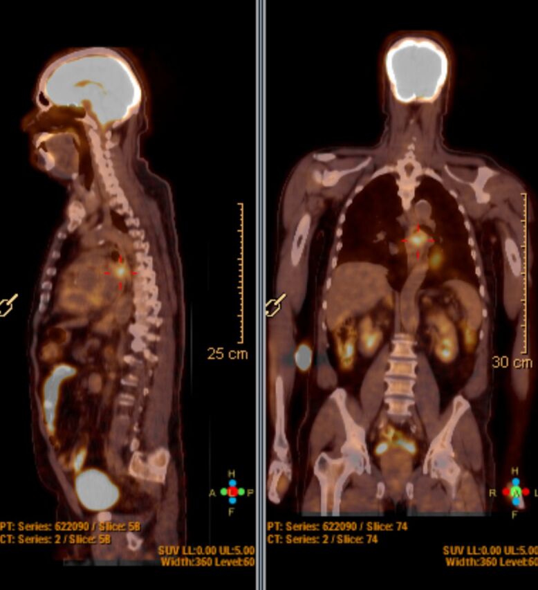
Gastrointestinal stromal tumours (GISTs) are the most common mesenchymal tumours of the gastrointestinal tract. Oesophageal GISTs are extremely uncommon, accounting for 0.7% of all GISTs, and their management is surrounded by some debate. We report a case of a 70-year-old man who was incidentally diagnosed with an oesophageal lesion on a 18F-fluorodeoxyglucose positron emission tomography. An endoscopic study revealed a non-obstructing 40 mm oesophageal lesion. Endoscopic ultrasound showed a well-circumscribed submucosal tumour on the middle oesophagus. Fine-needle aspiration was positive for CD117 and the overall features were of a GIST. After an initial thoracoscopic approach, the tumour was completely enucleated through a thoracotomy incision. The patient experienced no surgical complications and was discharged on day 4. Histopathology and immunohistochemical staining confirmed a low-risk GIST.

Keywords: cancer intervention; cardiothoracic surgery; general surgery; oesophageal cancer; surgical oncology.


Conflict of interest statement

Competing interests: None declared.

Figures

Figure 1
Figure 1
Coronal and sagittal view of an FDG-PET showing a high-uptake FDG oesophageal lesion. FDG-PET, fluorodeoxyglucose positron emission tomography.
Figure 2
Figure 2
Axial view of an FDG-PET showing a high-uptake FDG oesophageal lesion. FDG-PET, fluorodeoxyglucose positron emission tomography.
Figure 3
Figure 3
Fine-needle aspiration under EUS of the oesophageal lesion. EUS, endoscopic ultrasound.
Figure 4
Figure 4
The enucleated tumour with 45 mm size.

References

    1. Robb WB, Bruyere E, Amielh D, et al. . Esophageal gastrointestinal stromal tumor: is tumoral enucleation a viable therapeutic option? Ann Surg 2015;261:117–24. 10.1097/SLA.0000000000000505 - DOI - PubMed
    1. Pence K, Correa AM, Chan E, et al. . Management of esophageal gastrointestinal stromal tumor: review of one hundred seven patients. Dis Esophagus 2017;30:1–5. 10.1093/dote/dox064 - DOI - PubMed
    1. Nemeth K, Williams C, Rashid M, et al. . Oesophageal GIST-A rare breed case report and review of the literature. Int J Surg Case Rep 2015;10:256–9. 10.1016/j.ijscr.2015.02.023 - DOI - PMC - PubMed
    1. Kang G, Kang Y, Kim K-H, et al. . Gastrointestinal stromal tumours of the oesophagus: a clinicopathological and molecular analysis of 27 cases. Histopathology 2017;71:805–12. 10.1111/his.13292 - DOI - PubMed
    1. Zhang F-B, Shi H-C, Shu Y-S, et al. . Diagnosis and surgical treatment of esophageal gastrointestinal stromal tumors. World J Gastroenterol 2015;21:5630–4. 10.3748/wjg.v21.i18.5630 - DOI - PMC - PubMed

Publication types

MeSH terms