Review
. 2021 Feb;28(2):427-438.
doi: 10.1038/s41418-020-00648-0. Epub 2020 Oct 31.

Ubiquitin signaling in cell cycle control and tumorigenesis

Affiliations
Review

Ubiquitin signaling in cell cycle control and tumorigenesis

Fabin Dang et al. Cell Death Differ. 2021 Feb.

Abstract

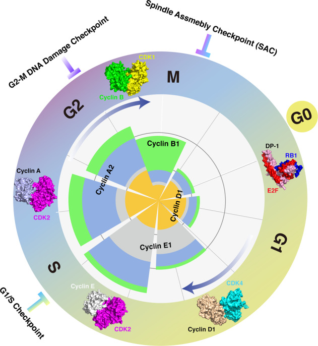
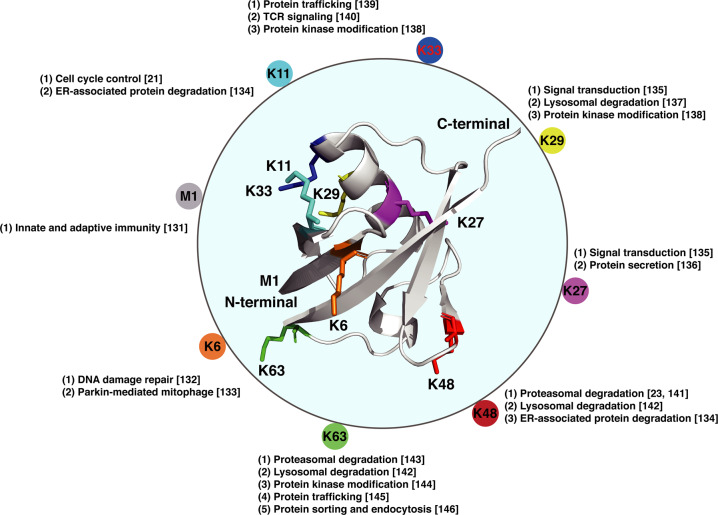
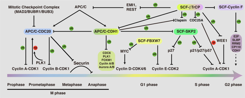
Cell cycle progression is a tightly regulated process by which DNA replicates and cell reproduces. The major driving force underlying cell cycle progression is the sequential activation of cyclin-dependent kinases (CDKs), which is achieved in part by the ubiquitin-mediated proteolysis of their cyclin partners and kinase inhibitors (CKIs). In eukaryotic cells, two families of E3 ubiquitin ligases, anaphase-promoting complex/cyclosome and Skp1-Cul1-F-box protein complex, are responsible for ubiquitination and proteasomal degradation of many of these CDK regulators, ensuring cell cycle progresses in a timely and precisely regulated manner. In the past couple of decades, accumulating evidence have demonstrated that the dysregulated cell cycle transition caused by inefficient proteolytic control leads to uncontrolled cell proliferation and finally results in tumorigenesis. Based upon this notion, targeting the E3 ubiquitin ligases involved in cell cycle regulation is expected to provide novel therapeutic strategies for cancer treatment. Thus, a better understanding of the diversity and complexity of ubiquitin signaling in cell cycle regulation will shed new light on the precise control of the cell cycle progression and guide anticancer drug development.


Conflict of interest statement

WW is a co-founder and consultant for the ReKindle Therapeutics. Other authors declare no competing financial interests.

Figures

Fig. 1
Fig. 1. Overview of the mammalian cell cycle.
The stages of the cell cycle are divided into four major phases: (1) G1 phase, also called the first gap phase. During the G1 phase, cells grow physically larger and duplicate cellular contents to prepare for the later steps; (2) S phase, cells synthesize a complete copy of DNA and duplicate the centrosome; (3) G2 phase, the second gap phase, cells grow more and prepare for mitosis; (4) M (mitotic) phase, during this phase, cells divide their copied DNA and cellular components, making two identical daughter cells. G0 phase is a quiescent stage that occurs outside of the cell cycle. During the G0 phase, cells are neither dividing nor preparing to divide. Sequential activation of Cyclin/CDKs drives cell cycle progression in a timely orchestrated manner. Briefly, Cyclin D1/CDK4 mainly functions in G1 phase to facilitate RB1 phosphorylation, releasing its suppression on E2F transcription factors; Cyclin E/CDK2 functions in S phase to control DNA replication; Cyclin A/CDK2 functions in later S phase to prepare the cell cycle entry into M phase; Cyclin B/CDK1 functions in M phase to be involved in regulation of chromatin separation. Additionally, three cell cycle checkpoints, G1/S checkpoint, G2-M DNA damage checkpoint, and spindle assembly checkpoint (SAC), are orchestrated to ensure the proper progression of the cell cycle. Protein structures of Cyclin/CDKs and RB1/E2F used here are as follows: RB1/E2F/DP (2AZE); Cyclin D1/CDK4 (2W9Z); Cyclin E/CDK2 (1W98); Cyclin A/CDK2 (6P3W); Cyclin B/CDK1 (4YC3).
Fig. 2
Fig. 2. A schematic diagram of the ubiquitination process.
Ubiquitination is an enzyme-mediated posttranslational modification by which the ubiquitin protein is attached to a substrate protein. This process involves three main steps: (1) activation step, the ubiquitin protein is activated by an E1 ubiquitin-activating enzyme, with ATP providing energy; (2) conjugation step, the ubiquitin protein is transferred from E1 to the active site of an E2 ubiquitin-conjugating enzyme; (3) ligation step, the ubiquitin protein is attached to the substrate (sub) with the catalyzation of an E3 ubiquitin ligase. Protein structures used here are as follows: Ub (1UBQ); E1–Ub complex (6DC6); E2–E3–Ub complex (4AP4).
Fig. 3
Fig. 3. Molecular structure of the ubiquitin molecule and linkage-dependent function of ubiquitination.
Ubiquitin is a small protein (8.6 kDa) that is expressed in all eukaryotic cells. There are eight amino acids (the N-terminal methionine M1 and seven lysine residues: K6, K11, K27, K29, K33, K48, and K63) that can serve as docking points for additional ubiquitin addition. The ubiquitination can be either a single ubiquitin protein (monoubiquitination) or a chain of ubiquitin (polyubiquitination). The variety of different modifications confers the diversity of linkage-dependent function of ubiquitination. Structure of ubiquitin is 1UBQ.
Fig. 4
Fig. 4. Function and regulation of the APC/C and SCF E3 ligases throughout the cell cycle.
APC/C and SCF E3 ligases serve as the two important types of E3 enzymes to regulate the cell cycle progression. Briefly, APC/C-CDC20 functions in prophase to metaphase to mediate the ubiquitination and proteasomal destruction of Cyclin A/B, Securin; APC/C-CDH1 functions from anaphase to G1 phase to modulate the protein stability of CDC6, PLK1, FOXM1, Cyclin A/B, and Aurora A/B, ensuring M phase progression and G1 phase maintenance. Four F-box proteins of SCF E3 ligase complex have been well-documented to function in regulation of the cell cycle progression: FBXW7, βTrCP, SKP2, and Cyclin F. FBXW7 functions largely as a tumor suppressor to mediate the protein destruction of MYC and Cyclin E. By contrast, SKP2 is believed to serve as an oncogene, which mediates the ubiquitination and degradation of CDK inhibitors, such as p21, p27, and p57. βTrCP is found to play a dual role in controlling CDK1 activity, turning it on by inducing Claspin and WEE1 degradation in G2 phase, and turning it off by inducing the degradation of CDC25A, EMI1, and REST. Cyclin F functions in G2 phase to restrict the activity of E2F, the synthesis of replicative histones (SLBP), and the levels of ribonucleotides (RRM2), as well as regulate centrosomal duplication (CP110). In addition, the APC/C and SCF E3 ligases control each other. For example, CDH1 mediates the degradation of SKP2 in G1 phase, while βTrCP and Cyclin F were reported to control the destruction of CDH1. Moreover, CDH1 is responsible for mediating CDC20 for proteasomal degradation, while the protein stability of Cyclin F was shown to be controlled by βTrCP.
Fig. 5
Fig. 5. Protein accumulation profiles of APC/C and SCF adapter proteins during the cell cycle.
A Western blot showing the protein abundance of APC/C and SCF adapter proteins over the course of the cell cycle. B Quantification of protein density showing the accumulation of protein levels of APC/C and SCF adapter proteins throughout the cell cycle.

References

    1. Weinberg RA. The Retinoblastoma protein and cell cycle control. Cell. 1995;81:323–30. doi: 10.1016/0092-8674(95)90385-2. - DOI - PubMed
    1. Zarkowska T, Mittnach S. Differential phosphorylation of the Retinoblastoma protein by G1/S cyclin-dependent kinases. J Biol Chem. 1997;272:12738–46.. doi: 10.1074/jbc.272.19.12738. - DOI - PubMed
    1. Hinds PW, Mittnacht S, Dulic V, Arnold A, Reed SI, Weinberg RA. Regulation of retinoblastoma protein functions by ectopic expression of human cyclins. Cell. 1992;70:993–1006. doi: 10.1016/0092-8674(92)90249-C. - DOI - PubMed
    1. Pagano M, Pepperkok R, Verde F, Ansorge W, Draetta G. Cyclin A is required at two points in the human cell cycle. EMBO J. 1992;11:961–71.. doi: 10.1002/j.1460-2075.1992.tb05135.x. - DOI - PMC - PubMed
    1. Petersen BO, Lukas J, Sørensen CS, Bartek J, Helin K. Phosphorylation of mammalian CDC6 by cyclin A/CDK2 regulates its subcellular localization. EMBO J. 1999;18:396–410. doi: 10.1093/emboj/18.2.396. - DOI - PMC - PubMed

Publication types

MeSH terms