. 2020 Nov 1;6(1):277.
doi: 10.1186/s40792-020-01057-8.

Reconstruction of the middle hepatic vein using a vein graft from the resected portion of the liver

Affiliations

Reconstruction of the middle hepatic vein using a vein graft from the resected portion of the liver

ShiWei Yang et al. Surg Case Rep. .

Abstract

Background: The middle hepatic veins are often infiltrated by intrahepatic cholangiocarcinoma. Reconstruction of the hepatic vein plays a critical role in preserving more of the residual liver volume and reducing the risk of postoperative liver failure in extreme hepatectomy. We here report a novel way to reconstruct middle hepatic vein by using vessel grafts from wasted liver.

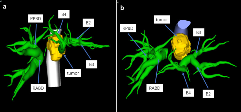
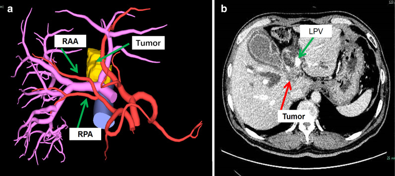
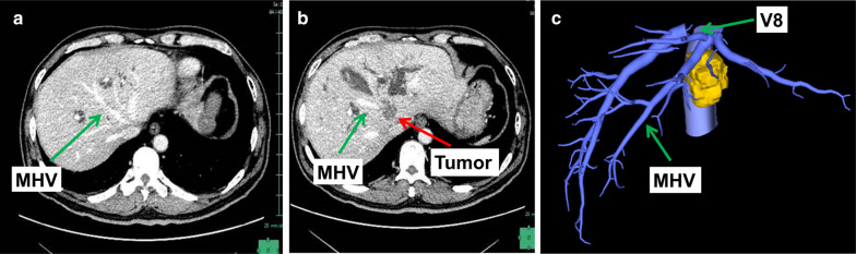
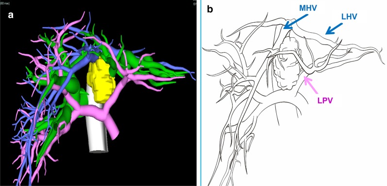
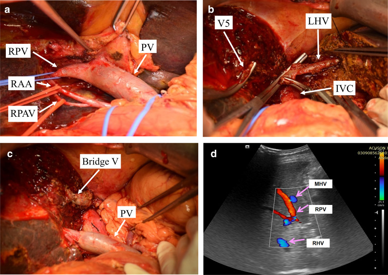
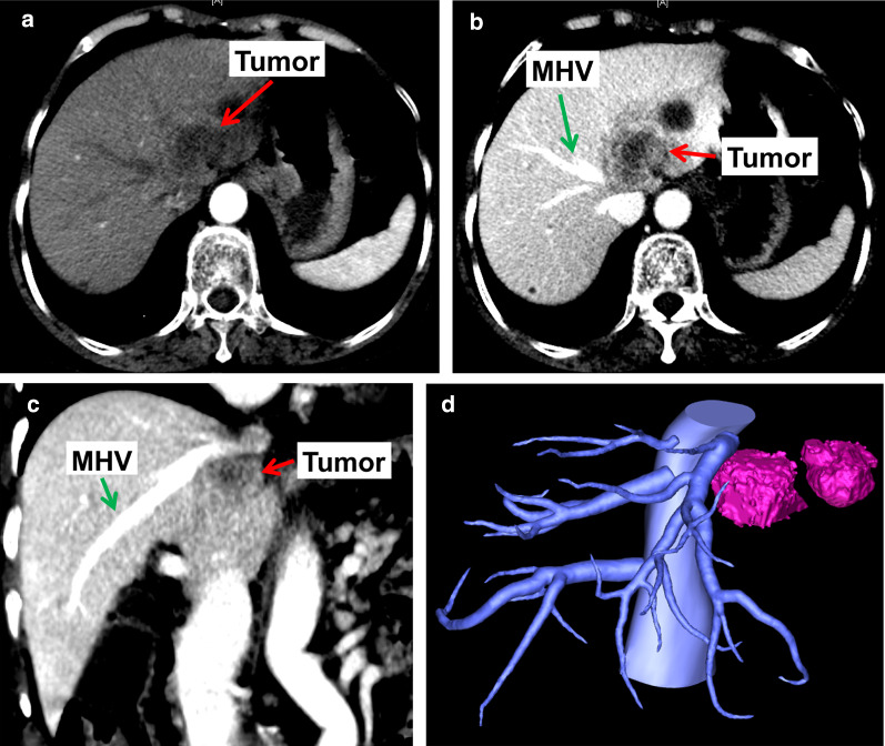
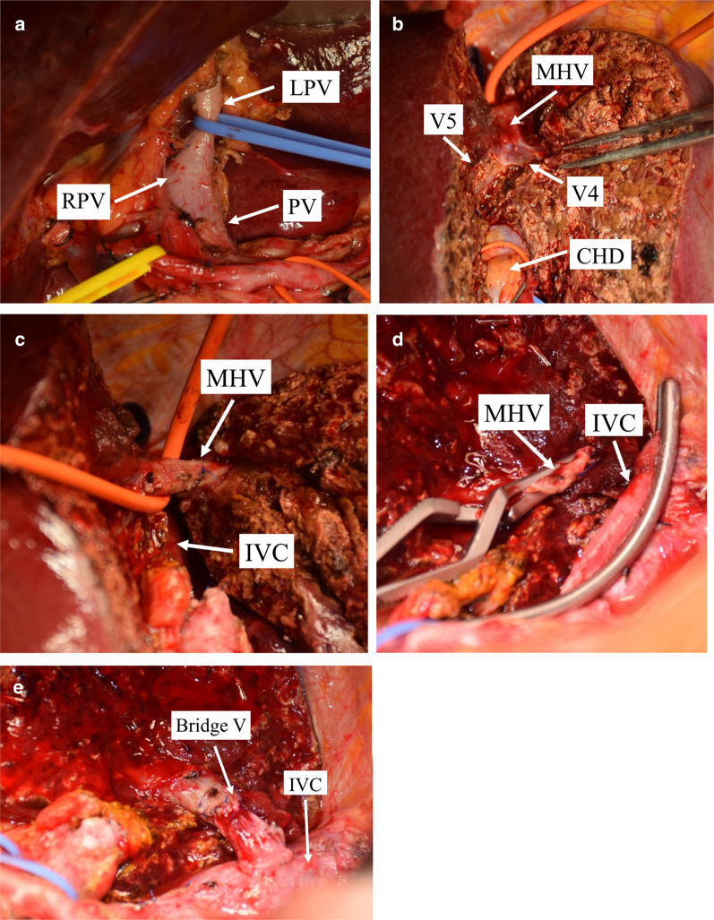
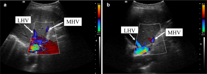
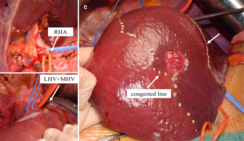
Case presentation: Case 1: A 64-year-old man was diagnosed with intrahepatic cholangiocarcinoma. The bifurcation and left branch of the portal vein were stenosed, and the root of the middle hepatic vein was infiltrated by the tumor. An extended left hepatectomy was performed, the portal vein was resected and reconstructed, and the middle hepatic vein was reconstructed by anastomosing the proximal left hepatic vein to the distal middle hepatic vein. Case 2: A 69-year-old woman was diagnosed with intrahepatic cholangiocarcinoma. The tumor was located in the left lobe of the liver and the left and middle hepatic veins were infiltrated by the tumor. An extended left hepatectomy was performed, and the left portal vein was used as a vein graft to reconstruct the middle hepatic vein. Both of the two patients' postoperative ultrasound showed vessel graft patency.

Conclusion: Using a vein graft from the resected portion of the liver to reconstruct the middle hepatic vein was a useful technique and showed good result.

Keywords: Autologous vein; Intrahepatic cholangiocarcinoma; Reconstruction of middle hepatic vein.


Conflict of interest statement

The authors declare that they have no competing interests.

Figures

Fig. 1
Fig. 1
Three-dimensional reconstruction of the tumor and bile duct shows the tumor was located at the hilar bile duct, and the left and right intrahepatic bile ducts were dilated. a Shows the relationships between bile duct with tumor in sagittal section. b Shows the relationships between bile duct with tumor in transverse section. B2 bile duct of segment 2, B3 bile duct of segment 3, B4 bile duct of segment 4, RABD bile duct of right anterior liver lobe, RPBD bile duct of right posterior liver lobe
Fig. 2
Fig. 2
Location of tumor and vessels: a the three-dimensional reconstruction of the tumor and artery, the right branch was closely related to the tumor, and the right hepatic artery was from the superior mesenteric artery and was not affected. b The CT scan which shows the tumor was located at the bifurcation of the portal vein, the left branch of the portal vein was compressed. RAA right anterior hepatic artery, RPA right posterior hepatic artery, LPV left portal vein
Fig. 3
Fig. 3
Relationship between tumor and hepatic veins: a and b are CT scan which shows that the tumor infiltrated the root of the middle hepatic vein, and the left hepatic vein was not infiltrated. c The three-dimensional reconstruction of the tumor and hepatic vein. MHV middle hepatic vein, V8 hepatic vein of segment 8
Fig. 4
Fig. 4
Fusion images of the tumor and bile ducts: a the three-dimensional reconstruction of vessels and tumor. b Shows the relationship between tumor with portal vein and hepatic vein. The tumor infiltrated the bifurcation of portal vein and left branch, bifurcation of hilar bile duct, and the middle hepatic vein. MHV middle hepatic vein, LHV left hepatic vein, LPV left portal vein
Fig. 5
Fig. 5
Three-dimensional reconstruction of the liver segments: a shows the area dominated by portal vein branch: S2: 99 mL (5.9%), S3: 74 mL (4.4%), S4: 237 mL (14.2%), S5: 186 mL (11.2%), S6: 182 mL (10.9%), S7: 409 mL (24.5%), S8: 479 mL (28.8%). b Shows the area dominated by hepatic vein branch: RHV: 630 mL (37.8%), LHV: 243 mL (14.6%), V8-1: 108 mL (6.5%), V8-2: 233 mL (14.0%), V5-1: 122 mL (7.3%), V5-2: 140 mL (8.4%), V4: 150 mL (9.0%). S2: segment 2, S3: segment 2, S4: segment 4, S5: segment 5, S6: segment 6, S7: segment 7, S8: segment 8, LHV: left hepatic vein, RHV: right hepatic vein, V4: hepatic vein of segment 4, V5-1: a branch of MHV which drained part one of segment 5, V5-2: a branch of MHV which drained part two of segment 5, V8-1: a branch of MHV which drained part one of segment 8, V8-2: a branch of MHV which drained part two of segment 8. MHV middle hepatic vein
Fig. 6
Fig. 6
Schematic diagram of the operation: a shows the location to remove MHV and RPV. b Shows the way to reconstruct MHV and RPV. The left hepatic vein was used for reconstruction of the middle hepatic vein, because it was far away from the tumor which ensured the R0 resection of the tumor and the entirety of hepatic blood inflow and outflow. MHV middle hepatic vein, LHV left hepatic vein, RPV right portal vein
Fig. 7
Fig. 7
Intraoperative hepatic vascular management: a skeletonization of right hepatic artery, portal vein. The bifurcation of portal vein was infiltrated by the tumor. b The root of the middle hepatic vein was infiltrated, after dissecting part of left hepatic vein from its root and rotated its direction, the main trunk of the middle hepatic vein was reconstructed with end to end anastomosis. c Reconstruction images of portal vein and hepatic vein. d Ultrasound on postoperative day 60 shows good blood flow in the right branch of the portal vein and middle hepatic vein. RPV right portal vein, PV portal vein, MHV middle hepatic vein, LHV left hepatic vein, RAA right anterior artery, RPA right posterior artery, IVC inferior vena cava, Bridge V bridge vein
Fig. 8
Fig. 8
The intrahepatic cholangiocarcinoma was located in root of hepatic vein, which infiltrated the middle hepatic vein and the left hepatic vein. a Shows the location of tumor in artery phase. b Shows the relationship between tumor and MHV in transverse section. c Shows the relationships between tumor and MHV in coronal section. d The three-dimensional reconstruction of tumor and hepatic vein. MHV middle hepatic vein.
Fig. 9
Fig. 9
a Shows the area dominated by portal vein branch. The area dominated by portal vein branch: S2 + S3 + S4: 352 mL (36.0%), S5 + S8: 459 mL (47.0%), S6 + S7: 146 mL (14.9%). b Shows the area dominated by hepatic vein branch. The area dominated by hepatic vein branch: V4-a: 76 mL (7.9%), V4-b: 214 mL (22.4%), V5: 154 mL (16.1%), V8-1: 30 mL (3.1%), V8-2: 45 mL (4.7%), V6: 168 mL (17.6%), V7-1: 60 mL (6.3%), V7-2: 150 mL (15.7%).S2: segment 2, S3: segment 2, S4: segment 4, S5: segment 5, S6: segment 6, S7: segment 7, S8: segment 8, V4-a: hepatic vein of segment 4a, V4-b: hepatic vein of segment 4b,V5: hepatic vein of segment 5, V6: hepatic vein of segment 6, V7-1: a branch of RHV which drained part one of segment 7, V7-2: a branch of RHV which drained part two of segment 7, V8-1: a branch of MHV which drained part one of segment 8, V8-2: a branch of MHV which drained part two of segment 8. MHV middle hepatic vein, RHV right hepatic vein
Fig. 10
Fig. 10
Intraoperative vascular operation: a dissected the first hepatic hilum, showing the left and right branches of the portal vein. b The trunk of middle hepatic vein and its branches. c Cut off the branch of the segment 4 originating from the middle hepatic vein. d The middle hepatic vein and the left hepatic vein were cut off from the root. The left liver lobe and the Spiegel lobe were removed to expose the inferior vena cava. e Using the left main branch of the portal vein as a vein graft to reconstruct the middle hepatic vein. LPV left portal vein, RPV right portal vein, PV portal vein, MHV middle hepatic vein, CHD common bile duct, IVC inferior vena cava, Bridge V bridge vein
Fig. 11
Fig. 11
Ultrasound on postoperative day 4 (a) and postoperative day 60 (b). MHV middle hepatic vein, LHV left hepatic vein
Fig. 12
Fig. 12
After blocking the right hepatic artery (a) and the middle and left hepatic veins (b), the hepatic congested area in segment 5 and segment 8 was revealed (c), indicating that this was the region drained by MHV. MHV middle hepatic vein, LHV left hepatic vein, RHA right hepatic artery

References

    1. Sozener U, Gulpinar K, Ozer Y, et al. Five right hepatic vein reconstructions using the autologous saphenous vein in the right lobe living-donor liver transplant: a case report. Exp Clin Transplant. 2014;12(2):159–161. - PubMed
    1. Sakamoto Y, Yamamoto J, Saiura A, et al. Reconstruction of hepatic or portal veins by use of newly customized great saphenous vein grafts. Langenbecks Arch Surg. 2004;389(2):110–113. doi: 10.1007/s00423-003-0452-9. - DOI - PubMed
    1. Shimagaki T, Yoshizumi T, Itoh S, et al. Liver resection with right hepatic vein reconstruction using the internal jugular vein: a case report. Surg Case Rep. 2016;2(1):132. doi: 10.1186/s40792-016-0258-y. - DOI - PMC - PubMed
    1. Del Campo C. Reconstruction of the hepatic and portal veins using a patch from the right ovarian vein. Am J Surg. 2000;179(1):77–78. doi: 10.1016/S0002-9610(99)00248-2. - DOI - PubMed
    1. Kubota K, Makuuchi M, Sugawara Y, et al. Reconstruction of the hepatic and portal veins using a patch graft from the right ovarian vein. Am J Surg. 1998;176(3):295–297. doi: 10.1016/S0002-9610(98)00149-4. - DOI - PubMed

LinkOut - more resources