. 2021 Apr;121(4):464-476.
doi: 10.1055/s-0040-1718760. Epub 2020 Nov 1.

Extracellular Histones Inhibit Fibrinolysis through Noncovalent and Covalent Interactions with Fibrin

Affiliations

Extracellular Histones Inhibit Fibrinolysis through Noncovalent and Covalent Interactions with Fibrin

Matthew Locke et al. Thromb Haemost. 2021 Apr.

Abstract

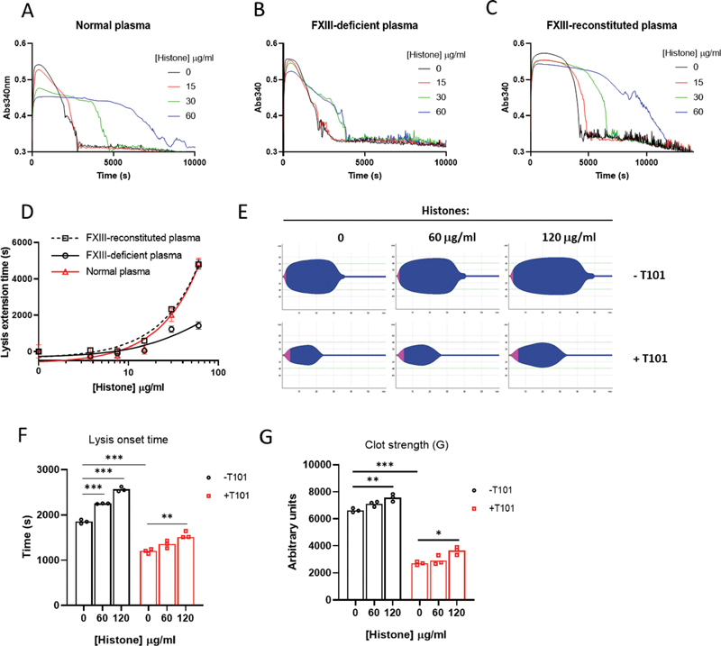
Histones released into circulation as neutrophil extracellular traps are causally implicated in the pathogenesis of arterial, venous, and microvascular thrombosis by promoting coagulation and enhancing clot stability. Histones induce structural changes in fibrin rendering it stronger and resistant to fibrinolysis. The current study extends these observations by defining the antifibrinolytic mechanisms of histones in purified, plasma, and whole blood systems. Although histones stimulated plasminogen activation in solution, they inhibited plasmin as competitive substrates. Protection of fibrin from plasmin digestion is enhanced by covalent incorporation of histones into fibrin, catalyzed by activated transglutaminase, coagulation factor FXIII (FXIIIa). All histone subtypes (H1, H2A, H2B, H3, and H4) were crosslinked to fibrin. A distinct, noncovalent mechanism explains histone-accelerated lateral aggregation of fibrin protofibrils, resulting in thicker fibers with higher mass-to-length ratios and in turn hampered fibrinolysis. However, histones were less effective at delaying fibrinolysis in the absence of FXIIIa activity. Therapeutic doses of low-molecular-weight heparin (LMWH) prevented covalent but not noncovalent histone-fibrin interactions and neutralized the effects of histones on fibrinolysis. This suggests an additional antithrombotic mechanism for LMWH beyond anticoagulation. In conclusion, for the first time we report that histones are crosslinked to fibrin by FXIIIa and promote fibrinolytic resistance which can be overcome by FXIIIa inhibitors and histone-binding heparinoids. These findings provide a rationale for targeting the FXIII-histone-fibrin axis to destabilize fibrin and prevent potentially thrombotic fibrin networks.


Conflict of interest statement

None declared.

Figures

Fig. 1
Fig. 1
Histones inhibit plasmin to delay fibrinolysis in a purified system. ( A ) tPA or plasmin (5 nM) was incubated with S-2288 or S-2251, respectively, and the indicated concentration of mixed histones. Initial rates were calculated from plots of absorbance (405 nm) versus time and expressed relative to the condition without added histone. Points represent means ± SEM from duplicate wells. ( B ) Plasmin activity was measured with increasing amounts of S-2251 in the presence of the indicated histones. The inset shows a Lineweaver–Burk plot to illustrate competitive inhibition of plasmin by histones. ( C ) Completed reactions from (A) for 240 μg/mL histones were analyzed by SDS-PAGE and Coomassie staining. H1 histone, core complex (H2A, H2B, H3, and H4), and digested histones are annotated. The control sample represents histones without tPA or plasmin incubation. ( D ) Plasmin was added to the surface of fibrin clots prepared by mixing fibrinogen, thrombin, and Ca 2+ with the indicated concentration of histone. Fibrinolysis was monitored by absorbance (405 nm) and turbidity values normalized as described in the Materials and Methods section. Lysis curves represent means of three experiments. ( E ) Fibrin clots with or without histones (240 μg/mL) were incubated with plasmin and solubilized at the indicated time points for reducing SDS-PAGE and Coomassie staining. The positions of fibrin(ogen) α, β, and γ chains and crosslinked variants (γ − γ dimer and α n -polymers), together with digestion products of γ − γ dimer (γ' − γ') and β chain (β′′), are annotated. Bands corresponding to the β chain ( F ), γ − γ dimer ( G ), and α n polymers ( H ) were quantitated and expressed relative to their starting intensities. Data represent mean ± SEM ( n  = 3). *** p  < 0.001 ( I ). Rates of plasminogen activation by tPA in the presence of increasing histones were measured by hydrolysis of S-2251 and calculated from plots of absorbance versus time squared. Points shown are means ± SEM from duplicate wells. ( J ) Internal clot lysis assays catalyzed by tPA were performed by clotting purified fibrinogen with thrombin in the presence of increasing histones, Ca 2+ , plasminogen, and tPA. Inhibition of fibrinolysis by histones is expressed as extension to 50% lysis times. Errors bars are ± SEM from duplicate wells. SEM, standard error of mean; tPA, tissue type-plasminogen activator.
Fig. 2
Fig. 2
Histones are crosslinked to fibrin in purified, plasma, and whole blood systems. ( A ) Purified fibrinogen was mixed with thrombin in the presence or absence of Ca 2+ , FXIIIa inhibitor T101 (200 μM), and increasing amounts of histones (30, 60, or 120 μg/mL). Insoluble fibrin was isolated from the clot by centrifugation and solubilized for analysis by SDS-PAGE and Coomassie staining ( bottom panel ) or western blotting with anti-histone H3 antibodies ( upper panel ). The positions of crosslinked and noncrosslinked fibrin(ogen) are annotated on the Coomassie gel. The expected positions of noncovalently bound, monomeric histone H3 (∼14 kDa) and high molecular weight crosslinked forms are annotated on the western blot. ( B ) Fibrin clots were prepared as in (A) in the presence of mixed histones (120 μg/mL) and clotting stopped at the indicated times. ( C ) Histones (240 μg/mL) and T101 were added to clotting plasma. After 2 hours, clots were dissolved in solubilization buffer and analyzed by western blot with antibodies specific for fibrin(ogen) and histone H3. The positions of crosslinked fibrin and histone are annotated. ( D ) FXIII-deficient plasma was incubated with histones (240 μg/mL) and clotted as in (A), with or without addition of purified FXIII (0.25–1.0 IU/mL). Crosslinked species were detected as in (A). ( E ) Whole blood was clotted in the presence of the indicated concentration of histone and/or T101 (200 μM) for 2 hours at room temperature. Clots were collected by centrifugation followed by homogenization/washing to obtain the isolated fibrin component, which was dissolved and analyzed by SDS-PAGE and Coomassie staining or western blotting. Fibrin–fibrin and histone–fibrin crosslinked products are annotated.
Fig. 3
Fig. 3
Histones accelerate fibrin polymerization and enhance clot structure independent of crosslinking to fibrin. ( A–C ) Purified fibrinogen was clotted by thrombin in the presence of the indicated amounts of histone in buffer containing Ca 2+ (A), without Ca 2+ (B), or containing T101 (200 μM) plus Ca 2+ (C). Fibrin formation was monitored by absorbance (405 nm). Curves are average of triplicate measurements. ( D ) Time to 50% clotting (half-maximum absorbance), ( E ) rates of lateral aggregation of protofibrils, and ( F ) fibrin fiber mass-to-length ratios calculated from turbidity measurements from A–C (see the Materials and Methods section and Supplementary Fig. S4 [available in the online version] for details). Rates in (E) are relative to clots without Ca 2+ and histones. Error bars in D–F represent 95% confidence intervals of the mean from three measurements. Statistical analysis was performed using one-way ANOVA and is relative to conditions without histones. * p  < 0.05, *** p  < 0.001. ANOVA, analysis of variance.
Fig. 4
Fig. 4
The antifibrinolytic potency of histones is enhanced by crosslinking to fibrin. ( A–C ) Normal plasma, FXIII-deficient plasma, or FXIII-deficient plasma supplemented with 1 IU/mL purified FXIIII were clotted in the presence of the indicated histone and 0.6 nM tPA. Clotting and lysis were monitored at 340 nm. Curves are averages of three experiments. ( D ) Extension to 50% lysis times by histones calculated from clot lysis curves in A–C. Error bars are ± SEM from three experiments. ( E ) Citrated blood was incubated with histones, tPA (2.5 nM), and T101 (200 μM), and clotting initiated by extrinsic pathway activation (EXTEM). Viscoelastic clot strength was monitored by rotational thromboelastometry (ROTEM) and is represented as clot firmness ( y -axis, arbitrary units) over time ( x -axis, minutes). ( F ) Lysis onset time (defined as the time taken for maximum clot firmness to decrease by 15%) and ( G ) clot strength (defined by G , shear strength) derived from the TEMograms in (E). Each bar indicates the mean from the three measurements represented by each dot. Statistical analysis was performed using one-way ANOVA. Asterisks represents difference from “zero histone” control ± T101. * p  < 0.05, ** p  < 0.01, *** p  < 0.001. ANOVA, analysis of variance; SEM, standard error of mean; tPA, tissue type-plasminogen activator.
Fig. 5
Fig. 5
Low-molecular-weight heparin (LMWH) prevents histone–fibrin crosslinking and neutralizes the antifibrinolytic potency of histones. ( A ) Fibrinogen was clotted by thrombin in the presence of the indicated concentration of histone and LMWH for 2 hours, followed by isolation of insoluble fibrin from the clot by centrifugation. Fibrin was solubilized and analyzed by SDS-PAGE and Coomassie staining or western blotting. ( B, C ) Raw clot lysis profiles of fibrinogen clotted with thrombin in the presence of plasminogen, tPA, Ca 2+ , and the indicated histone without (B) or with 1 IU/mL LMWH (C). ( D ) Extension to 50% clot lysis times by histones calculated from clot lysis curves in B and C. ( E ) Maximum turbidity of clot lysis curves in B and C. Error bars in D and E represent ± SEM from duplicate measurements. SEM, standard error of mean.
Fig. 6
Fig. 6
Summary of histone effects on fibrin stability and fibrinolysis. Histones are associated with fibrin through noncovalent interactions and covalent crosslinking catalyzed by FXIIIa. Noncovalent histone–fibrin interactions increase lateral aggregation of fibrin protofibrils and increase fibrin-fiber thickness, giving the fibrin network increased mechanical stability. Histones stimulate plasminogen activation by tPA in the absence of fibrin, possibly by providing a surface for colocalization of reactants through histone–lysine plasminogen–kringle interactions. However, histones competitively inhibit plasmin to delay fibrinolysis, being digested in the process, and protect fibrin from degradation. Crosslinked histones are more effective at inhibiting fibrinolysis than noncovalently bound histones and increase the biochemical stability of fibrin. Blocking histone–fibrin crosslinking with histone-binding heparinoids, or FXIIIa inhibitors, improves lysis of clots containing histones. This suggests targeting the FXIII–histone–fibrin axis could be effective in destabilizing clots containing histones to prevent thrombosis. tPA, tissue type-plasminogen activator.

Similar articles

Cited by

References

    1. Chen R, Kang R, Fan X G, Tang D. Release and activity of histone in diseases. Cell Death Dis. 2014;5:e1370. - PMC - PubMed
    1. Brinkmann V, Reichard U, Goosmann C.Neutrophil extracellular traps kill bacteria Science 2004303(5663):1532–1535. - PubMed
    1. Engelmann B, Massberg S. Thrombosis as an intravascular effector of innate immunity. Nat Rev Immunol. 2013;13(01):34–45. - PubMed
    1. Gould T J, Lysov Z, Liaw P C. Extracellular DNA and histones: double-edged swords in immunothrombosis. J Thromb Haemost. 2015;13 01:S82–S91. - PubMed
    1. Fuchs T A, Brill A, Duerschmied D. Extracellular DNA traps promote thrombosis. Proc Natl Acad Sci U S A. 2010;107(36):15880–15885. - PMC - PubMed